

- Discussion:
- first performed by Darrach in 1911 in New York City.
- Indications:
- for relief of pain following distal RU disruption and/or RU arthritis;
- for symptomatic malunion of Colle's frx in elderly patients, especially when stiffness is present;
- procedure is generally performed on elderly patients w/ low functional demands;
- young pts w/ symptomatic instability of distal RU after Colle's frx may have better results w/ distal radial osteotomy w/ restoration of length & alignment, or when this is not possible then consider hemi-resection arthroplasty;
- Technique:
- dorsal approach to the wrist
- longitudinal incision is made over distal ulna w/ care to preserve the attachments of the TFCC to the carpi;
- alternatively, approach distal ulna thru the interval between the ECU and FCU;
- carefully place Homan retractors around the ulna, w/ care to avoid injury to the ulnar artery and nerve;
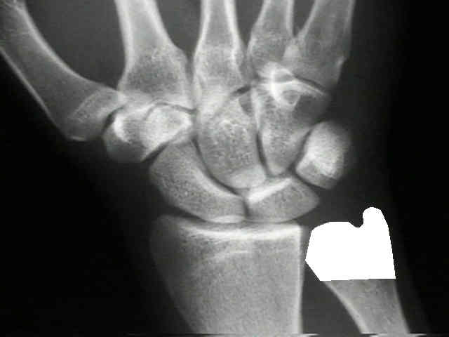
- excise of distal 1-2 cm of ulna (just proximal to the sigmoid notch);
- some surgeons attempt to minimize the amount of bone resected, inorder not to destabilize the distal ulna, and to maintain attachments of pronator quadratus;
- the least amount of bone is excised which is sufficient to restore full motion;
- ulnar styloid process and its attachment to the ulnar collateral ligament may be preserved;
- bevel any sharp edges;
- remnants of the TFCC are opposed to the wrist capsule and the radius;
- remnants of the wrist capsule are anchored to the distal ulna;
- if the ECU is volarly subluxated, it should be relocated and anchored distally over the carpus;
- Stabilizing Procedures:
- if the distal ulna appears unstable, it may be stabilized w/ a distally based strip of ECU tendon pass through a drill hole at the end of the bone;
- the tendon is sutured to itself with the wrist in ulnar deviation;
- ECU/pronator quadratus arthroplasty:
- distally based ECU tendon is attached to the dital ulna and helps prevent radial-ulnar impingement;
- the pronator quadratus arthroplasty helps retard dorsal translation;
- standard approach between the ECU and the FCU tendons;
- pronator quadratus is dissected off of its insertion on the ulna (palmar medial side);
- the pronator is mobilized, and is then transferred over the dorsal surface of the ulna;
- medullary canal of the distal ulna is reamed with a burr, and a drill hole is prepared approximately 1.5 cm proximal to the distal end ofthe ulna;
- harvest one half of the ECU tendon, leaving it attached distally;
- pass the free half of the ECU through the medullary canal of the distal ulna and then back out of the hole made in the side of the ulna;
- flex the elbow and keep the forearm in neutral rotation;
- pass 2 divergent K wires from the ulna to the radius while maintaining a desired amount of radioulnar distance;
- these wires allow soft tissue healing and are left in place for 6 weeks;
- Complications:
- procedure does not produce uniformly good results, especially in younger patients;
- instability of the distal ulnar shaft;
- painful subluxation of the ECU over the transected end of the ulna;
- palmar subluxation or ulnar translation of the carpi;
- radio-ulnar impingement (convergence);
- occurs between stump of the ulna and the ulnar side of radius and is usually accompanied by instability;
- impingement is worsened w/ power grip, and the pronator quadratus seems to be the major deforming force;
- severity of the convergence correlates w/ the amount of resected bone;
- often causes symptoms, but as reported by McKee and Richards, convergence can occur w/o symptoms;
- when this complication occurs, consider the stabilization procedures, described previously;
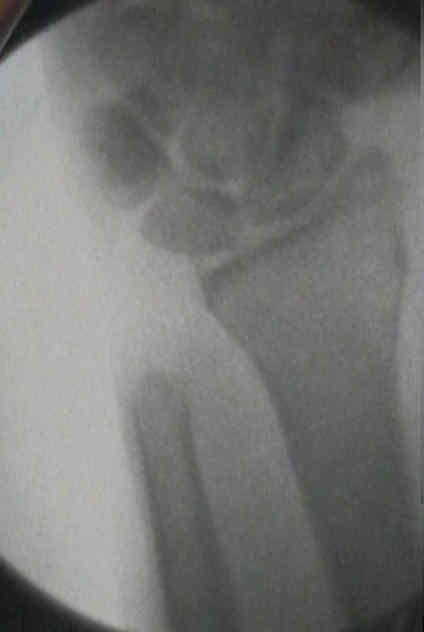
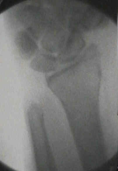
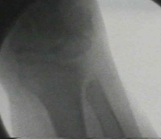
- case example:
- this patient had convergency type pain, which was thought to be due in part to a contracture of the prontator quadratus
- Dynamic radio-ulnar convergence after the Darrach procedure.
Anterior dislocation of the head of the ulna. Darrach W. Ann Surg. 1912;56:802-803.
Habitual forward dislocation of the head of ulna. Darrach W. Ann Surg. 1913;57:928-930.
Partial excision of lower shaft of ulna for deformity following Colle's fracture. Darrach W. Ann Surg. 1913;57:764-765.
Poor results of Darrach's procedure after wrist injuries.
Results of extensor carpi ulnaris tenodesis in the rheumatoid wrist undergoing a distal ulnar excision.
Rupture of digital extensor tendons following distal ulnar resection.
A modified extensor carpi ulnaris tenodesis with the Darrach procedure.
Extensor carpi ulnaris and flexor carpi ulnaris tenodesis of the unstable distal ulna.
Salvage of the failed Darrach procedure.
History of hand surgery
William Darrach, MD: His Life and His Contribution to Hand Surgery.