

- Discussion:
- coalition bewteen talus & calcaneus may be difficult to detect on xray
- cartilaginous coalition between the talus and calcaneus in the area of the middle facet may also be detected by an irregularity of the joint surfaces of the talus and calcaneus;
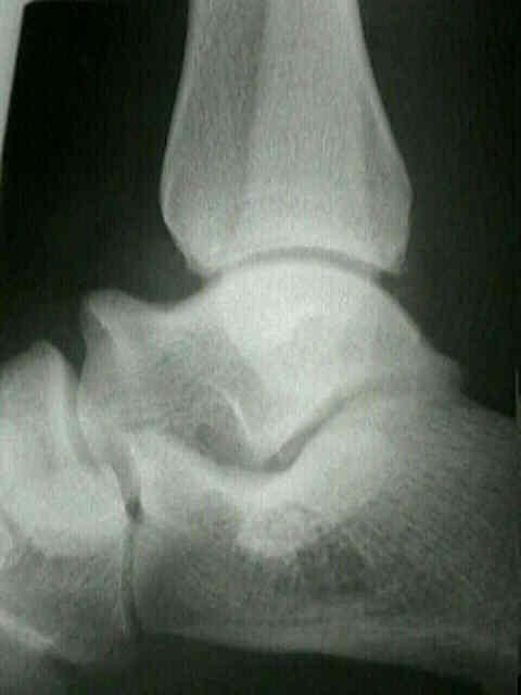
- anterior facet is not seen on the axial x-ray, and tomography is required to visualized it properly;
- Lateral View:
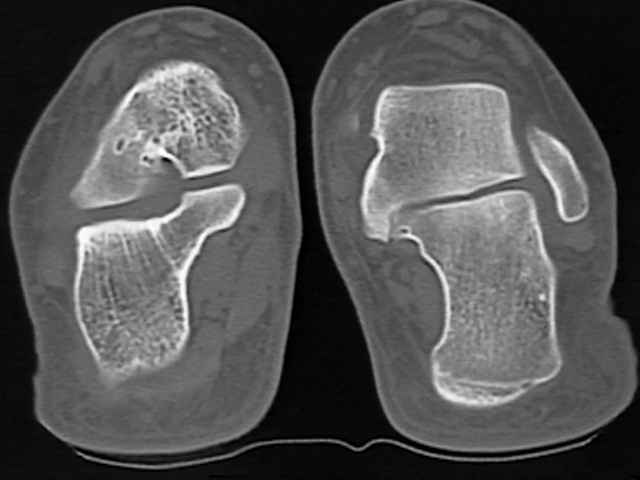
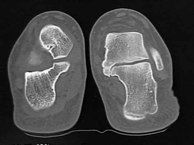
- CT Scan:
- 5 mm cuts are acceptable but 3 mm cuts are more optimal;
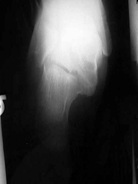
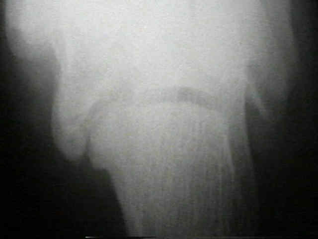
- Harris View:
- since normal posterior & middle facet are in parallel planes at approx 45 deg to sole of foot, these two areas can be identified on an Axial view (described by Korvin in 1933);
- effectiveness of Harris View for visualizing subtalar joint is enhanced by measuring angle of posterior facet on lateral view & then adjusting Harris view to this inclination;
- normally the middle facet is perpendicular to the axis of the tibia;
- angulation of middle facet by > 20 deg off horizontal is consistent w/ coalition, even if the joint space is open;
- posterior & middle facet of calcaneus, which articulate w/ talus, are nl in parallel planes & are usually best visualized on axial radiographs at 45 deg;
- in talocalcaneal coalition, angle of middle facet may vary;
- anterior facet is in a different plane and is not seen axial views because it is obsurred by head of the talus
Evaluation of tarsal coalition by computed tomography.
Computerized tomography (CT) scanning technique for the hindfoot.