

Discussion
pathoanatomy
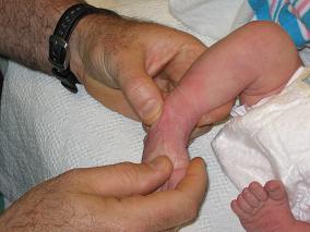
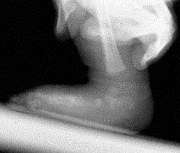
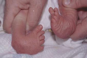
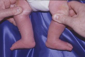
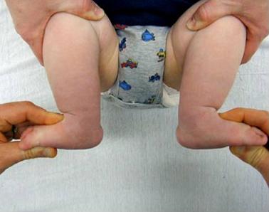
- whole foot is in extreme supination, however, the fore part of the foot is pronated with respect to the hindfoot, as a result of the cavus deformity (the first metatarsal is more plantar flexed than the fifth metatarsal);
- navicular and the cuboid are rotated medially in relation to the talus, and are held in adduction and inversion by contracted ligaments and tendons;
- tibial-navicular interval:
- distance between the medial malleolus and the tuberosity of the navicular;
- shorter intervals indicate worse deformity;
- degree of resistance of navicular to be moved away from medial malleolus correlates with severity of deformity.
- note that in severe clubfoot, complete reduction of the extreme medial displacement of the navicular may not be possible by manipulation;
timing
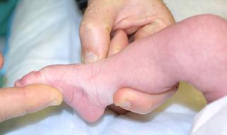
- casting begins in the first week of life inorder to take advantage of the initial elasticity of contracted ligaments, joint capsules and tendons;
- improvements from manipulation are maintained by immobilizing the foot in a plaster cast for five to seven days;
- within the first 2-3 months, the surgeon attempts 5-6 manipulation and cast applications;
- children who present for treatment after four or five months old may require operative correction because ligaments become stiffer;
- total duration of treatment should be less than three months;
- 6-8 toe-to-groin plaster casts, changed weekly after manipulation and worn for 7-10 weeks, should be sufficient to obtain maximum correction possible;
Sequence of Correction: (Ponseti)
correction of cavus
- cavus deformity must be corrected prior to correcting the other deformities;
- forefoot is supinated and the first metatarsal is dorsiflexed;
- this reverses the contracted forefoot pronation;
- pronation of the foot will worsen the deformity and will increase the cavus;
- an attempt to correct the inversion of the foot by forcible pronation of anterior part of the foot increases the cavus deformity as first metatarsal is plantar-flexed further;
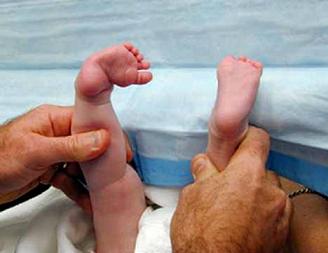
correction of adduction and heel varus
- goal is to abduct the supinated foot under the talus;
- again, forceful pronation of the foot is avoided since it increases the cavus deformity, causes mid foot break down and does not address the varus heel deformity;
- talus is rotated laterally so that the foot abducts underneath the talus which is fixed in the ankle mortice;
- this causes lateral rotation of navicular, together w/ cuboid & anterior aspect of calcaneus, w/o pronation of foot;
- to correct the varus and adduction, the foot in supination is abducted while counterpressure is applied with the thumb against the head of the talus;
- foot is abducted in flexion and slight supination to stretch the medial tarsal ligaments, while counter pressure applied on the lateral aspect of the head of the talus;
- this allows the calcaneus to abduct under the talus which correction of the heel varus;
- heel must not be touched during this manipulation;
- foot is abducted in flexion and slight supination to stretch the medial tarsal ligaments, while counter pressure applied on the lateral aspect of the head of the talus;
- calcaneus abducts by rotating and sliding under the talus;
- noted that the calcaneus can evert only when it is abducted (laterally rotated) under the talus.
- as the calcaneus abducts it simultaneously extends and everts which corrects the heel varus;
- note that the calcaneus cannot evert unless it is abducted;
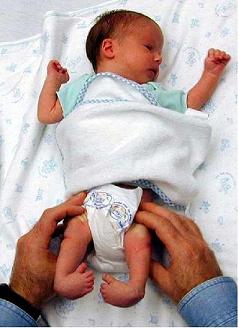
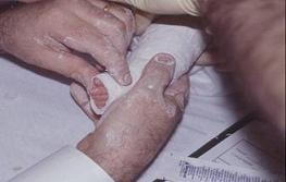
- casting involves a toe-to-groin plaster cast w/ knee flexed 90 degrees and the foot in maximum external rotation;
- maintenance of correction of varus deformity of hind part of foot which requires external rotation of foot distal to talus;
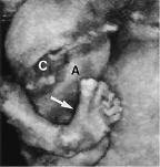
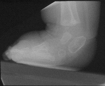
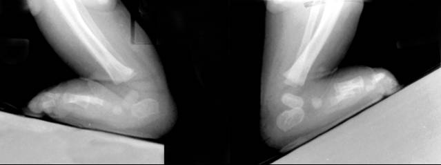
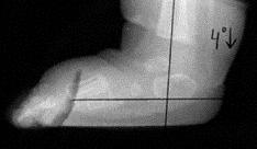
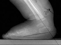
- radiographs may be taken at this point inorder to confirm that the talonavicular joint is reduced, prior to managing equinus;
- cautions:
- avoid forced external rotation of the foot to correct adduction while the calcaneus is in varus;
- this causes a posterior displacement of the lateral malleolus by externally rotating the talus in the ankle mortice.
- avoid abducting the foot against pressure at the calcaneocuboid joint the abduction of the calcaneus is blocked, thereby interfering with correction of the heel varus
- avoid forced external rotation of the foot to correct adduction while the calcaneus is in varus;
correction of equinus:
- equinus is corrected last, by dorsiflexion of foot w/ heel in valgus angulation;
- if foot is dorsiflexed prior to correction of the hindfoot varus, rocker bottom foot may be created;
- equinus is corrected by dorsiflexing the fully abducted foot;
- correction entails stretching of the tight posterior capsules and ligaments of ankle and subtalar joints and the tendo achillis;
- lateral x-ray are helpful in assessing quality of cast correction;
- percutaneous tenotomy of the achillis tendon:
- may be necessary inorder to avoid rocker bottom deformity;
- dorsiflexion of ankle to > 10 to 15 degrees is rarely possible because of talar and calcaneal malformations and tight ligaments;
- cautions:
- care should be taken not to cause a rocker-bottom deformity, which can occur when dorsiflexion of foot is attempted w/ pressure under metatarsals rather than under the mid-part of foot, particularly when varus deformity of heel has not been corrected;
- do not to exert excessive upward force on metatarsals, because this can result in midfoot break (rocker-bottom deformity);
- ref: Radiographic Evaluation of Idiopathic Clubfeet Undergoing Ponseti Treatment
Complications
- increased cavus deformity;
- rocker-bottom deformity;
- longitudinal breach
- flattening of the proximal surface of the talus
- lateral rotation of the ankle
- increased stiffness of the ligaments and joints;
- recurrence:
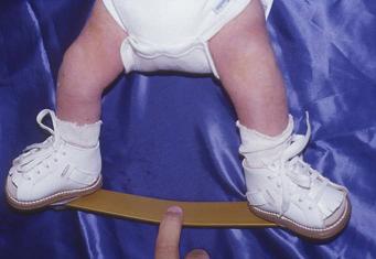
- Ponseti advocates use of shoes attached to a bar in external rotation for three months full-time and at night for 2-4 years
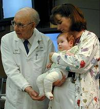
Masters in Surgery

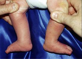
Motivation for Ponseti Method: Current Surgical Methods:
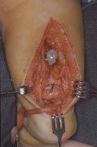
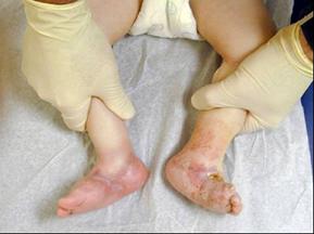
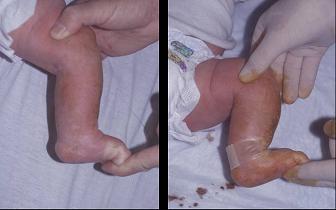
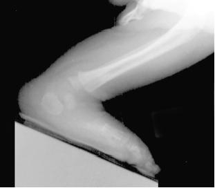
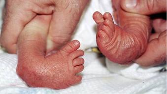
Example of Wound Breakdown following Surgery:
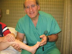
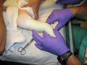
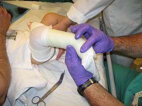
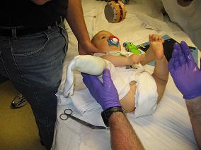
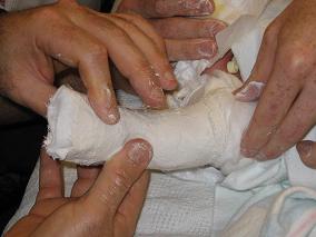
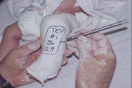
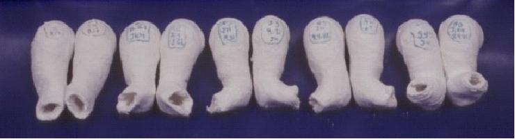
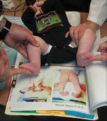
Casting Considerations for the Ponseti Technique: 1
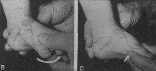
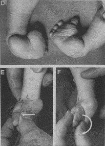
Casting Considerations for the Ponseti Technique: 2
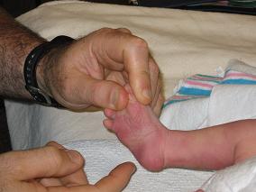
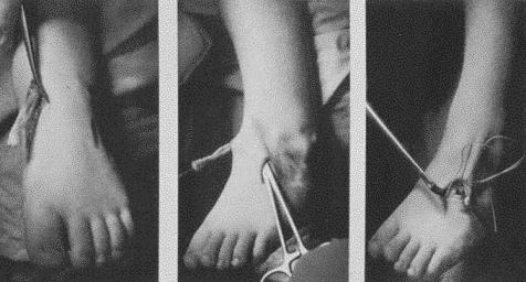
Achilles Tenotomy:
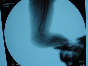
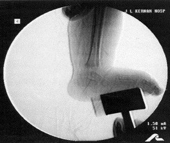
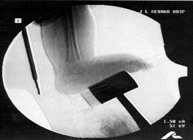
Use of x-rays to assist with achilles tentomy:
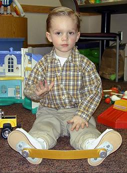
Dennis Brown Bar:
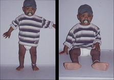
Outcomes:

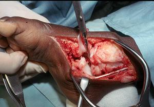
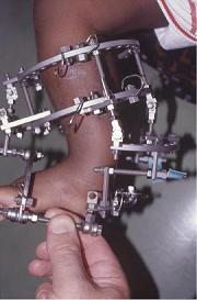
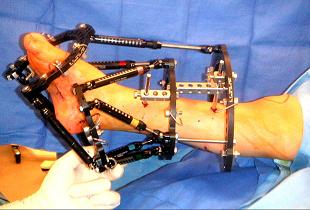
Surgical Case Example: Ilizarov Method
Compliance:
References
- Ponseti Versus Traditional Methods of Casting for Idiopathic Clubfoot
- Long-Term Comparative Results in Patients with Congenital Clubfoot Treated with Two Different Protocols.
- Long term results of treatment of congenital clubfoot.
- Congenital club foot: the results of treatment.
- Factors Predictive of Outcome After Use of the Ponseti Method for the Treatment of Idiopathic Clubfeet.
- The Ponseti method of treatment for congenital clubfoot: importance of maximal forefoot supination in initial casting.
- Ponseti versus traditional methods of casting for idiopathic clubfoot.
- Results of manipulation of idiopathic clubfoot deformity in Malawi by orthopaedic clinical officers using the ponseti method: a realistic alternative for the developing world?
- Results of an accelerated Ponseti protocol for clubfoot.
- Early Clubfoot Recurrence After Use of the Ponseti Method in a New Zealand Population
- Calf Circumference Discrepancies in Patients With Unilateral Clubfoot: Ponseti Versus Surgical Release
- Ponseti Method Compared with Surgical Treatment of Clubfoot. A Prospective Comparison