

- Discussion:
- fibular plate can be positioned posteriorly as an antiglide plate to resist proximal migration and rotation of distal fragement;
- the antiglide plate is chiefly indicated for Weber B fractures;
- advantages:
- distal screws obtain better purchase, since they engage a thicker part of distal fibula, and engage 2 cortices without risk of joint penetration;
- biomechanically this contruct is stronger than lateral plate fixation, especially in osteoporotic bone;
- loss of fixation (w/ subsequent malunion or nonunion) is rare due to the stability of the construct;
- in elderly patients, posterior plate position may be fixation of choice, since it is biomechanically stronger;
- posterior plate provides better fixation w/ posterior comminution;
- essentially no risk of intra-articular screw insertion;
- less risk of wound slough than with direct lateral approach, and there is essentially no risk of symptomatic palpable screws;
- posterior incision allows access to the posterior malleolus, when direct fixation is required;
- disadvantages: may irritate peroneal tendons in a minority of patients, but this often spontaneously resolves in 4-8 weeks;
- peroneal tendon subluxation should not be a problem is the tendon sheath is left intact;
- Technique:
- patient can be positioned supine on a bean bag;
- lateral malleolus frx:
- patient is already on the bean bag and can placed in the lateral position;
- the foot can be place on a padded Mayo stand for support;
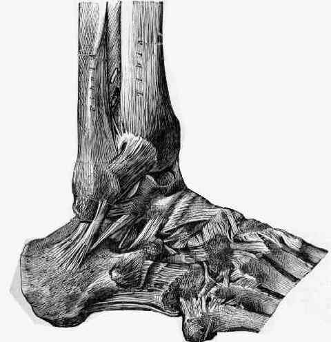
- surgical approach:
- peroneal tendons are retracted posteriorly, providing access to the fibula;
- care is taken not to enter the peroneal tendon sheath;
- some of proximal retinaculum may need to be released to expose distal fibula;
- periosteum is clear off the posterior surface of the fibula;
- frx reduction:
- idealy an anatomic should be achieved prior to plate application;
- reduction can be held with a single K wire or lag screw;
- if lag screw is inserted it must be applied in a direct posterior direction so as not to interfere with plate application;
- plate application:
- one-third tubular plate is applied to posterior surface of the fibula;
- the classic technique used a 4-hole plate, but in a study by Ostrum R (1996), most of the fractures were fixed w/ a 6 hole plate;
- because the posterior surface of fibula is straight, contouring of the plate is usually not necessary;
- due to the lateral bow of the fibula, the plate may have to be placed posterolaterally so that the distal portion of the plate does not veer medially off the fibula;
- Posterior Plating of Displaced Weber B Fibula Fractures
- proximal screw insertion:
- first 3.5 mm cortical screw is placed proximally,
through the plate, 2 mm above the posterior frx line;
- if an antomic reduction has already been achieved, the proximal screw can be tightened down;
- if an anatomic reduction has not been achieved, then do not fully tighten the screw;
- plate helps prevent proximal gliding of the distal fragment;
- apply bone holding clamps to both frx fragments, and pull the frx out to length, and apply slight internally rotate the distal fragment;
- reduction should be achieved w/ this maneuver;
- at this point the proximal screw is tightened down;
- remaining proximal 3.5 mm cortical screws are inserted;
- lag screw:
- lag screw is then inserted (posterior to anterior) thru the first plate hole which is distal to the posterior frx line;
- the screw must be angle slightly proximally inorder to be perpendicular to the frx site;
- as Schaffer and Monoli (1987) emphasize, lag screws do not significantly improve the biomechanical characterics of the antiglide plate, but they can improve the fracture reduction;
- because the posterior cortex is thin, the lag screw must be inserted through the plate (which serves as solid posterior cortex);
- because the screw is directed away from the peroneal tendons, there is no negative consequence of leaving the screw slightly long;
- if possible, a second lag screw can be inserted using the same technique;
- The antiglide plate for distal fibular fixation. A biomechanical comparison with fixation with a lateral plate
- distal screws:
- technically the more distal screws are not necessary, but 4.0 mm cancellous screws may be inserted at the surgeon's discretion;
- because there is no risk of joint penetration, longer screws can be used (upto 24 mm) and get a better hold on bone;
- medial malleolus fracture:
- 4.0 mm cancellous bone screws, or 4.0-4.5 mm cannulated screws are used for fixation of medial malleolus & posterolateral tibial fragment;
- alternatively use K wires, 1.6 mm diameter, and 1.2 mm cerclage wires for tension band wiring of the medial malleolus;
- exploration of ankle joint:
- need to look for osteochondral fragments;
- after exposure of frx & anterior surface of fibula, joint is explored;
- this should be repeated during the approach to the lateral malleolus;
- Post Op:
- if a syndesmotic screw has not been inserted, patients can be given the option at POD 14, of full weight bearing in a short leg cast, or TDWB w/ a plastic CAM walker (which can be taken off for bathing);
- Case Examples:
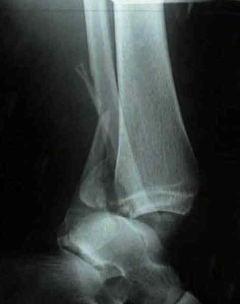
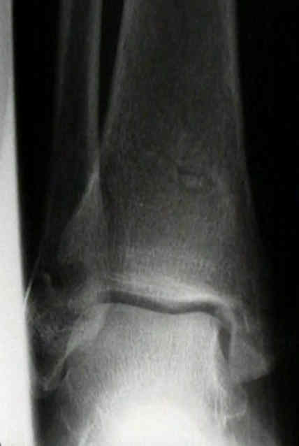
- this 40-year-old patient sustained a trimalleolar fracture from a fall;
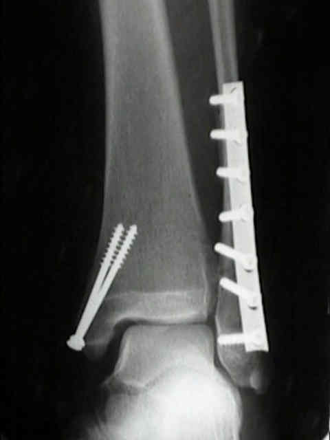
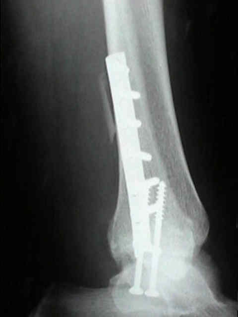
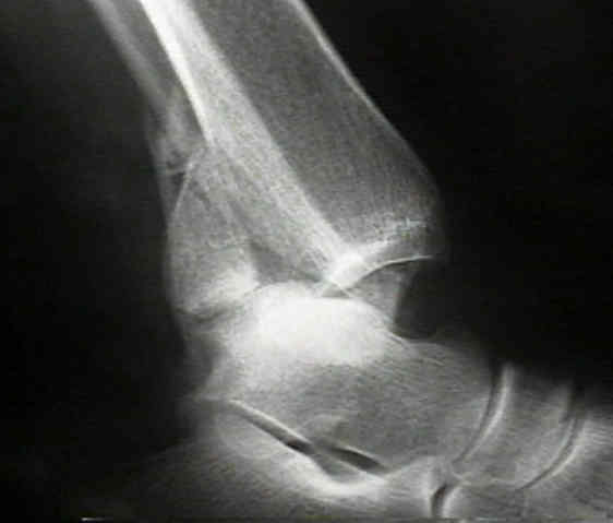
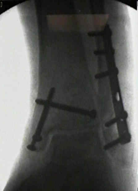
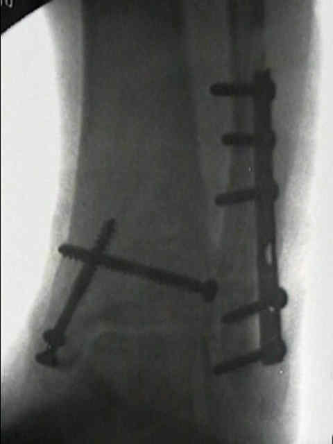
- the incision was made directly posterior, which allowed easy access to the posterior malleolar fracture by dissecting behind the FHL;
- the outline of the fracture was easily palpated thru the incision, which allowed insertion of the cannulated screw w/o flouroscopy;
- due to the anatomic lateral bow of the distal fibula, a 6 hole one third tubular plate could not be placed on the posterior surface of the fibula (the distal portion of the plate was not centered on the bone due to the lateral bow);
- therefore the plate was placed on the posterolateral surface of the fibula
The dorsal antiglide plate in the treatment of Danis-Weber type-B fractures of the distal fibula.