

- See: Arterial supply to Femur
- Embryology of Proximal Femur: 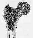
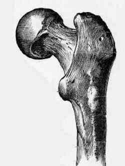
- upper end consists of head, neck, & greater & lesser trochanters at junction of the neck with the body;
- develops from 4 separate ossification centers;
- head forms roughly two-thirds of sphere whose surface is articular except for fovea capitis femoris where ligament of head is attached;
- greater trochanter is large prominence projecting upward from shaft on lateral aspect of junction of neck & body of femur;
- lesser trochanter is protuberance on posteromedial side;
- intertrochanteric crest, extends between two trochanters;
- wide, rough intertrochanteric line stretches from greater to lesser trochanter on the anterior side of the femur;
- calcar femorale
- Femoral Neck Angle:
- neck extends inferolaterally from head to meet shaft of femur at angle of about 125 deg;
- angle varies w/ age, stature, & width of pelvis, being less in adult, in persons with short limbs, and in women;
- when this angle is > 135 deg, condition is known as coxa valga;
- when < 120 deg, coxa vara;
- femoral neck is not parallel to frontal plane or plane of femur;
- instead, head is located anterior to midline of shaft of femur & is thus said to be anteverted;
- functionally this results in internal rotation of femoral shaft, so that Ipt w/ increased anteversion may walk w/ intoeing gait;
- in adult, neck - shaft angle is approx between 5-15 deg;
- when > 15 deg, increased femoral anteversion is present;
- when < 5deg, condition is termed femoral retroversion;
- references:
- The anatomic basis of femoral component design.
- Anteversion of the Femur and Acetabulum
- Femoral Shaft:
- femur is essentially a tubular structure;
- shaft of femur is slightly twisted and curved w/ convexity forward, partially accounting for fullness of the anterior thigh;
- it is most nearly cylindrical in its middle third;
- above & below it is sl flattened anteroposteriorly & widened, esp toward lower end;
- it flares posteriorly along linea aspera, where its cortical thickness is greatest;
- linea aspera, on back of middle third has two lips that diverge;
- at or near upper end of linea aspera is nutrient foramen;
- linea aspera serves as a site of attachment for the fascia;
- proximal & distal metaphyseal widening of the tube in subtrochanteric and supracondylar regions of bone results in stress concentrations at these levels;
- most prominent feature of femoral shaft is its anterior bow or antecurvature;
- wide individual variations exist in magnitude of this bow;
- normal physiologic bow is often increased in certain pathologic dzs, such as fibrous dysplasia & Paget's dz;
- clinical significance of antecurvature of the shaft has long been appreciatted;
- most modern IM nails are prebent, to accommadate the bow;
- straight, stiff implants used in early years of femoral nailing straightened shaft, leaving posterior gap at fracture site;
- straight nails also resulted in frx in frx comminution and occassionally even perforation of anterior cortex
Clinical determination of femoral anteversion. A comparison with established techniques.
The anatomy and functional axes of the femur.
Vascularised fibular grafts for reconstruction of the femur.
The arterial supply of the developing proximal end of the human femur.