

- See: Open Acromioplasty
- Discussion and Indications:
- objective good to excellent results may be achieved in over 70% of patients, and subjective satisfactory results are achieved in over 90% of patients;
- shoulder arthroscopy is indicated in cases of impingement syndrome and questionable rotator cuff tear repair, since the shoulder scope will be able to determine the status of the cuff, and an arthroscopic acromioplasty can be performed if a cuff tear is not present;
- outcomes:
- in the study by Spangehl MJ, et al, the authors evaluated whether arthroscopic acromioplasty is equivalent or superior to open acromioplasty, in a prospective, randomized, controlled, blinded clinical trial;
- Arthroscopic versus open acromioplasty: a prospective, randomized, blinded study
- indications:
- young athletic patient w/ stage II impingement and strong desire to return to spots;
- as reported by Tibone, et al (1985), only 43% of 33 patients were able to return to sports after open acromioplasty;
- in contrast, Altchek, et al (1990), reported that 76% of 33 atheletically inclined patients were able to return to sports;
- the patients that were unable to return to sports, usually had inferior labral pathology, indicating occult instability;
- average recovery time is 4 months;
- cautions: beware that impingement in younger patients may be due to occult anterior instability;
- Shoulder impingement syndrome in athletes treated by an anterior acromioplasty
- Arthroscopic acromioplasty. Technique and results.
- older patient w/ impingement and massive rotator cuff tear;
- in these patients, the goal should be relief of pain and not improvement in function (under the assumption that the cuff tear cannot be repaired);
- this subgroup of patients is also more likely to have degenerative lesions in other joints, which makes a quick rehabilitation essential (e.g., DJD of the contralateral hip would make use of a cane difficult);
- relative contra-indications:
- young to middle aged adult w/ significant weakness due to mid-sized (1-3 cm) rotator cuff tear, in which case an open acromioplasty might be more appropriate;
- type III acromion:
- as Rockwood as pointed out, acromioplasty (either open or arthroscopic) necessitates that the inferior and anterior attachments of the deltoid to the acromion must be released, leaving only the superior deltoid as an attachment;
- w/ a type III acromion, the deltoid stripping may especially large, and therefore the surgeon should consider open acromionplaty with reattachment of the deltoid to the anterior aspect of the acromial edge;
- os acromiale:
- may instead require open reduction and internal fixation and decompression;
- internal rotation contracture:
- limitation of external rotation worsens impingement syndrome since the greater tuberosity cannot clear the acromion as the arm is elevated;
- logically, by improving the internal rotation contracture, the impingement syndrome will be relieved;
- greater tuberosity osteophyte:
- if radiographs reveal a prominent greater tuberosity osteophyte (which is often co-existent w/ rotator cuff tear), then removal of the osteophyte is carried out by open means;
- Radiographs
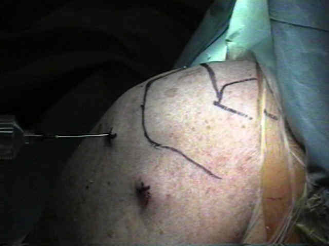
- Positioning:
- beach chair position is comfortable for both the patient and the surgeon and allows the option of converting to an open procedure (if necessary);
- patients need to be seated with the torso raised to 70 deg with a foled sheet placed on the medial border of the affected scapula (to maximize posterior exposure)
- simultaneously palpate posterolateral corner and anterior of the acromion and note the "arthoscopic plane";
- if this plane is sloped too steeply, then the surgeon will have to hold the arthroscope in an awkward position (w/ the scope pointing upwards);
- to remedy this problem, increase the patient's sitting angle which will bring the arthroscopic plane down to a more horizontal level;
- complications: hypoglossal nerve injury;
- reference:
Hypoglossal nerve palsy after arthroscopy of the shoulder and open operation with the patient in the beach-chair position. A case report.
- Portal Placement:
- generally arthroscopy of the shoulder joint is carried out prior to arthroscopic acromioplasty, and therefore, standard anterior and posterior arthroscopic portals are established;
- a self sealing cannula is usually placed through the anterior portal;
- precautions:
- prior to inserting instruments into the subacromial space it is helpful to prevent subacromial bleeding (or limit it before it occurs);
- pre-inject the subacromial space with an epinephrine containing solution;
- consider adding 1/2 ampule of epipnephrine to each 3 liter bag of NS;
- ensure that multiple fluid bags are ready;
- ensure that intra-articular cautery is ready;
- once the arthroscopic examination of the shoulder is completed, the arthroscope (posterior portal) is pull out of the shoulder joint and is driven into the subacromial space;
- if this portal has been placed too inferior, visualization of the subacromial joint may be difficult;
- anterior portal:
- the arthroscope is driven anteriorly until the tip can be palpated near the anterior portal;
- drive the arthroscope thru the anterior portal and then pull the scope back out of its cannula;
- the arthroscopic cannula serves as a positioning guide for the anterior cannula which is then driven into the subacromial space;
- a 5.5 mm shaver is placed into the anterior portal cannula and an inital arthroscopic bursectomy is carried out;
- one adequate visualization of the subacromial space has been established, the lateral portal is established;
- lateral portal: (see portal placement);
- used as the main instrument portal (acromioplasty and bursal debridement);
- lateral portal is inserted in the usual manner with care that its placement will allow full triangulation of the undersurface of the anterior acromion;
- Plan to Control Bleeding:
- pre-inject the subacromial space with 30 cc of 1:300,000 epinephrine solution;
- if possible, have anesthesia lower the patient's blood pressure;
- ensure that reserve fluid bags are present;
- arthroscopic techniques:
- when the posterior subacromial portal is established, bluntly sweep the trocar across the undersurface of the acromion which helps to remove the bursal attachments;
- minimize the use of the shaver to clear off the bursa, since this may stir up early bleeding;
- use cautery or the arthrocare wand to carefully define the undersurface landmarks of the acromion;
- be careful not to disturb the deltoid fascia which lies below the acromion since this is guaranteed to stir up bleeding;
- air injection:
- if bleeding obstructs the visualization, then shut off inflow and suction out the fluid;
- inject 100 cc of air through a syringe;
- bleeding will then usually stop spontaneously;
- Arthroscopic Decompressions:
- the posterior portal is used to view the subacromial space, the lateral portal is used for instrumentation, and the anterior portal is used for either outflow or inflow;
- a 6.5 mm cannula is inserted thru the anterior and lateral portals to accomadate the inflow and instrumentation;
- the first step usually involves a limited bursectomy using the 5.5 mm full radius shaver;
- it is essential to limit the bursectomy to the region around the anterior acromion, inorder to limit bleeding;
- establish landmarks:
- the anterior portal serves as a landmark for the AC joint;
- undersurface of acromion is covered by periosteal layer & layer of coracoacromial ligament, which extends under acromion (this covering is quite thick and extensive);
- the entire undersurface of the antero-lateral acromion is cleared of soft tissue using intra-articular cautery inorder to limit bleeding;
- take care not to stray into the deltoid fibers, since this will stir up bleeding and will ruin the case;
- once the bursal tissue is removed from the undersurface of the acromion, cautery is then used to remove bursal tissue and the deltoid attachements on the anterior acromial surface;
- note that bleeding from branch of thoracoacromial artery can easily occur when sectioning the coracoacromial ligament;

- Acromioplasty:
- arthroscopic impingement test:
- following removal of the inflamed bursal tissue an arthroscopic impingement test should bes performed;
- place the arthroscope through the lateral arthroscopic portal and then flex the shoulder (move back and forth from the scapular plane to straight forward);
- note any impingement between the humeral head and the acromion (following the acromioplasty no impingement should be observed and the subacromial space should be maintained);
- as has been described by Rockwood et al a two step acromioplasty is necessary;
- anterior acromioplasty:
- ideally, the portion of the acromion which extends anterior to the anterior edge of the clavicle should be removed (this is often about 5 mm and can be estimated from the 30 deg caudal tilt view);
- place a 5 mm burr on the cuff, just below the acromial tip;
- this gives an indication of how much space is present, and when the acromioplasty is finished, the burr is again used to judge the amount of space that is present;
- use the burr to remove the anterior 5 mm of the acromion, completing the most lateral side prior to removing the medial side of the acromioplasty;
- once the lateral half of the anterior acromion has been removed, it is easy to judge how much of the remaining acromion needs to be removed;
- inferior acromioplasty: (second stage acromioplasty)
- use a burr to create a centering hole 2 mm deep in the undersurface of the acromion, which is located 5 mm posterior the anterior acromial edge and which is located midway between the AC joint and the lateral aspect of the acromion;
- this centering hole is used to help plane the inferior acromioplasty;
- in some cases, it is helpful to switch the arthroscope to the lateral portal and place the burr in the posterior portal;
- Bursal Debridemnt:
- the bursa is debrided after the acromioplasty, since it tends to bleed more (disrupting the case);
- take special when debriding around the AC joint since bleeding is especially troublesome;
- rotate the arm internally and externally to expose different parts of the bursa;
- methodically sweep the shaver along the bursa;
- Controversies:
- distal clavicle excision::
- in the study by Fischer BW, et al (1999), the authors studied the effect of violation of the AC joint during arthroscopic acromioplasty;
- the authors found that patients that either had no violation of the AC joint or patients that had complete distal clavicle resection had no postoperative sequelae in reference to the AC joint;
- in contrast, 14 of the 36 shoulders (39%) with a documented AC joint violation and a partial distal clavicle resection developed acromioclavicular joint symptoms at an average of 8.4 months;
- the authors recommend an all or nothing approach to the AC joint (i.e., avoid partial distal clavicle resection);
- the authors recommend that if the AC joint must be violated to perform an adequate decompression of the subacromial space, complete resection of the distal clavicle should be performed, even if the radiographs show no preoperative degenerative changes;
- the authors theorize that partial distal clavicle excision increases AC joint motion in all planes, especially rotation and superior translation, after co-planing of the inferior surface;
- these authors proposed that the increased mobility could lead to acromioclavicular joint symptoms in a previously asymptomatic joint or accelerate symptoms in a joint with mild symptoms before surgery;
- ref: Incidence of acromioclavicular joint complications after arthroscopic subacromial decompression.
- rotator cuff tear tear:
- as pointed out by Gartsman et al, in a group of patients with partial thickness rotator tears, there was 33 satisfactory and 7 unsatisfactory results;
- as pointed out by Gartsman et al, in a group of patients with full thickness rotator tears, there was 14 satisfactory and eleven satisfactory results;
- in this same group, 6 out of 7 patients that underwent subsequent rotator cuff repair had a satisfactory result;
- from Altchek et al, 6 of 10 patients w/ a full thickness RTC, had a good or excellent result;
- these patients should generally expect some pain relief but no improvement in function from the arthroscopic acromioplasty;
- posterior capsular release:
- may be indicated in selected patients, w/ limited internal rotation and w/ thickened posterior capsule who demonstrate persistent impingement (as seen arthroscopically);
- technique involves arthroscopic visualization from the anterior portal and release of the posterior capsule off of the glenoid using cautery;
- reference: Recognition and Treatment of Refractory Posterior Capsular Contracture of the Shoulder
- Post Op:
- average time for full recovery is about 4 months

Arthroscopic subacromial decompression versus open acromioplasty. A two-year follow-up study.
Arthroscopic acromioplasty for lesions of the rotator cuff.
Arthroscopic acromioplasty. Technique and results.
Arthroscopic debridement versus open repair for rotator cuff tears. A prospective cohort study.
Partial thickness rotator cuff tears: results of arthroscopic treatment.
Arthroscopic treatment of massive rotator cuff tears. Clinical results and biomechanical rationale.
Rotator cuff pathology. Arthroscopic assessment and treatment.
Arthroscopic subacromial decompression: results in advanced impingement syndrome.
Arthroscopic treatment of massive rotator cuff tears. Clinical results and biomechanical rationale.
Shoulder impingement syndrome in athletes treated by an anterior acromioplasty.