- See: 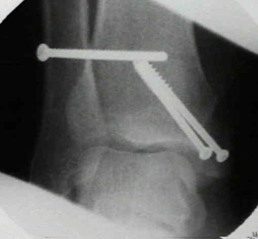 syndesmotic sprain
- Anatomy:
- syndesmosis is made up of anterior-inferior tibiofibular ligament, interosseous ligament, and
posterior-inferior fibular ligaments, inferior transverse tibiofibular ligament, and interosseous ligament;
- these stabilize the mortise by opposing the fibula in the fibular notch (incisura fibularis tibiae);
- section of the anterior tibiofibular ligament results in diastasis of 2.3 mm;
- section of anterior tibiofibular ligament and interosseous ligament will result in diastasis of 4.5 mm;
- when all 3 ligaments are sectioned, diastasis measures 7.3 mm;
- syndesmotic injuries are unusual in displaced Weber B fractures;
- in the anatomic study by Snedden MH and Shea JP, the authors noted that the interosseous ligament, may have a variable
attachment on fibula, differing between specimens in its distance above the synovial reflection or joint line;
- low fibula frx would disrupt interosseous ligament, can explain anatomic basis for infrequent diastasis in these ankle frxs;
- ref: Diastasis With Low Distal Fibula Fractures: An Anatomic Rationale.
- Injury Patterns:
- isolated syndesmotic injury:
- syndesmotic injury & fibular frx;
- w/ syndesmosis & fibula disruption, talus can shift laterally 2 to 3 mm, even w/ deep deltoid ligament intact;
- syndesmotic injury + medial injury:
- > 3 mm displacement indicates that either the deep deltoid ligament or medial malleolus must be disrupted;
- if medial malleolus is frxed & deltoid ligament is intact, rigid fixation of fibula & tibia should make
syndesmosis fixation unnecessary;
- posterior malleolus fracture
- fixation of posterior malleolar fractures will make syndesmotic reduction and fixation easier;
- Objective Diagnosis of Syndesmotic Injury and Indications for Syndesmotic Fixation:
- Surgical Treatment Options:
- reduction of syndesmosis (theory and surgical technique)
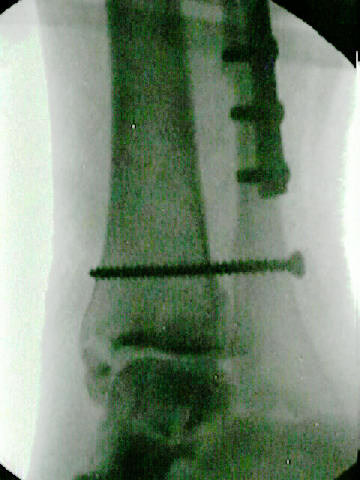
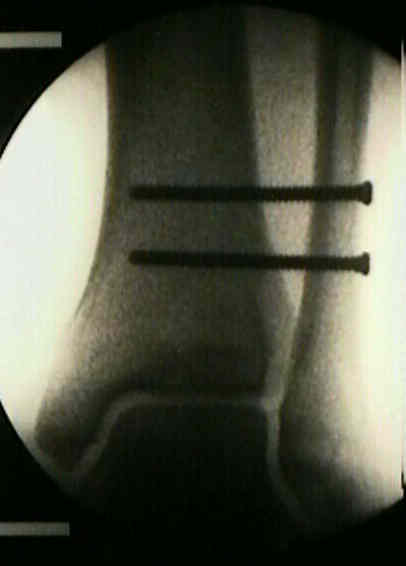
- fixation techniques:
- screw insertion technique for snydesmotic fixation
- k wire fixation:
- two 1.5 mm K wires can be inserted obliquely across the distal tibio-fibular syndesmosis;
- is a less rigid form of fixation, which allows more physiologic ankle function, and does not require early HW removal;
- deltoid ligament repair:
- ref: Deltoid Ligament Repair Versus Syndesmotic Fixation in Bimalleolar Equivalent Ankle Fractures.
- suture fixation (w/ suture button):
- involves creation of two small drill holes through fibula and tibia (separated 7-10 mm) above the ankle syndesmosis
(between 2-5 cm), through which is passed a single No 5 Ethibond suture to form a loop;
- the suture is tied over the fibula, securing the fibula to the tibia;
- advantages: there is no need for hardware removal and nor is there risk of hardware failure;
- in the study by Miller RS, et al (1999), the suture technique showed similar strength characteristics to tricortical screw
fixation techniques;
- references:
- Comparison of tricortical screw fixation versus a modified suture construct for fixation of ankle syndesmosis injury:
- Repair of the tibiofibular syndesmosis with a flexible implant.
- Comparison of a Novel FiberWire-Button Construct versus Metallic Screw Fixation in a Syndesmotic Injury Model
- Randomized Trial Comparing Suture Button with Single Syndesmotic Screw for Syndesmosis Injury
Long-term Results After Ankle Syndesmosis Injuries.
Examination and Repair of the AITFL in Transmalleolar Fractures.
Outcome after fixation of ankle fractures with an injury to the syndesmosis: the effect of the syndesmosis screw.
Mechanical considerations for the syndesmosis screw. A cadaver study.
Distal tibial fracture post syndesmotic screw removal: an adverse complication
The management of acute distal tibio-fibular syndesmotic injuries: Results of a nationwide survey.
Operative treatment of syndesmotic disruptions without use of a syndesmotic screw: a prospective clinical study.