

- Discussion:
- TKR: effects of malrotation
- patellar effects:
- malrotation of femoral component may lead to patellofemoral dislocation or subluxation;
- slight external rotation of femoral component on femur places patellofemoral groove more laterally, enhances patellofemoral stability (external
rotation moves the femoral component further lateral to the tibial tubercle);
- increased external rotation of AP cutting jig may also be necessary in patients with a severe fixed valgus deformity;
- consequences of too much external rotation
- when the femoral component is externally rotated, the proximal patellar track is lateralized but distally the groove is meidialized.
- so as the knee is more fully flexed, the patella it is medialized and can cause maltracking;
- An Alternative to the Consequences Associated with External Rotation of the Femoral Component in Total Knee Arthroplasty
- functional effects:
- femoral component of greater than 10°, regardless of the tibial component alignment, has negative effect on function during a
simulated squatting motion;
- large internal alignments of the femoral and tibial components place a greater demand on the quadriceps
- internal rotation of the femoral components generate higher forces on the MCL, causing stiffness
- ref: The biomechanical effects of variability in femoral and tibial component rotational alignment during TKR in a simulated oxford rig.
- piano sign
- if the femoral component has been placed in some external rotation, expect to see the piano sign from above (the lateral side will have a
nice flush cut, where as the medial side will appear undercut - resulting in the shape of a grand piano.)
- even though the piano sign is considered to be an indication of a properly performed anterior cut (proper external rotation),
the consequences is that anteromedially, the implant will be off the bone and there will be less mechanical fixation;
- references:
- Variations of the 'grand-piano sign' during total knee replacement. A computer-simulation study.
- Grand Piano Sign,” a Marker for Proper Femoral Component Rotation During Total Knee Arthroplasty


- AP Axis Method / Whitesides: method of choice
- Posterior Condyles as a Rotational Guide:
- generally the most reliable guide is the posterior condyles;
- w/ normal femoral anatomy, a slight relative external rotation (3 deg) of a line along the posterior aspect of the femoral condyles will
orient the AP cuts perpendicular to the resected tibial surface;
- the medial condyle extends below the epicondylar axis further than the lateral condyle and therefore, more of the medial condyle will be resected than the lateral;
- pitfalls:
- this technique, however, requires special care in any knee that has a large AP asymmetry in the femoral condyles;
- in such cases rotational alignment only w/ respect to posterior condyles may result in significant undercutting of the anterior cortex
laterally, severely notching the cortex;
- when the posterior condyles are not available as a rotational reference (such as in AVN or severe valgus deformity), the epicondyles &
form of the trochlea as seen in the skyline view can be used;
- varus deformity and effect on posterior condylar angle: (tibial varus deformity);
- in 60 consecutive TKR done in 52 patients w/ DJD and varus or neutral tibiofemoral alignment, posterior condylar angle average 3.98° (range, 0°-9°);
- 18 knees had a posterior condylar angle value less than 3° whereas 27 knees had a posterior condylar angle value of 5° or greater;
- final rotational alignment of the femoral component was set parallel to the transepicondylar axis;
- only one of these 60 knees required a lateral retinacular release for proper patellar tracking during the knee arthroplasty;
- as tibial varus joint line obliquity increased, there was tendency for transepicondylar axis to be rotated more externally relative to post condylar axis;
- hence, use of posterior condylar axis as a rotational reference is inappropriate in many knees w/ arthritis w/ varus or neutral tibiofemoral alignment;
- in particular, varus tibial joint line obliquity of more than 4 deg increases the likelihood of femoral component
malrotation when the posterior femoral condyles are used to reference femoral component rotation;
- ref: Varus Tibial Joint Line Obliquity. A Potential Cause of Femoral Component Malrotation.
- valgus deformity:
- w/ valgus deformity, there is defficiency of the of the posterior femoral condyle, which can lead to accidental internal rotation if the
typical 3 deg posterior condylar reference is used;
 Wheeless Method:
- initially use whatever method seems appropriate and apply the AP cutting guide;
- apply the extramedullary proximal tibial cutting guide, with great care to meticulously align the guide in the AP and lateral planes;
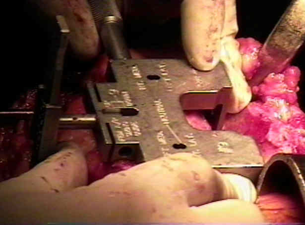
- with the knee flexed 90 deg, look at the flexion gap between the posterior edge of the sizing guide and adjust rotation
so that the posterior edge is parallel with the tibial cutting guide;
- often the fat pad and the patella tendon interfere with visualization so repeat process with AP cutting guide;
- apply AP cutting guide and assess whether it is parallel to the tibial cutting guide;
- check leg postion: make sure that the leg is straight up and not sagging to the side;
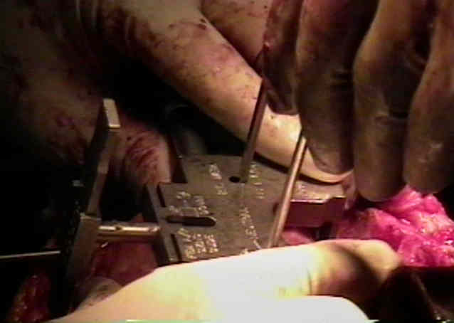
- if there is a flexion trapezoidal space, remove the guide, then use a drill bit to mill the guide holes (one up and one down) to
adjust rotation, and then reapply the AP guide;
- theoretiically no cuts should be made until there is a perfectly rectangular flexion gap;
- Transepicondylar Axis Method:
- connects the lateral epicondylar prominence and the medial sulcus of the medial epicondyle;
- medial sulcus is a more accurate landmark than the medial prominence;
- site of insertion of the deep part of the MCL;
- this method is probably most indicated in revision TKR;
- can be accurate method, but can be difficult to use do to overlying soft tissues;
- when measured carefully, the epicondylar axis most often will place the AP cutting guide in 4 deg of external rotation (which is ideal);
- note, however, that it may be common to error by placing the AP cutting guide in too much external rotation;
- in the report by Miller MC, et al., the authors evaluated the validity of this rotational landmark and its effect on the patellofemoral and tibiofemoral articulations.
- TKR was done in 11 knees from cadavers;
- knees were tested with various femoral component rotations from 5° internal rotation to 5° external rotation referenced to the epicondylar
axis and to the posterior femoral condyles;
- knees were actively ranged from 0° to 100° by a force on the quadriceps tendon in an Oxford knee simulator;
- femoral component rotation parallel to epicondylar axis resulted in best patellar tracking and minimized patellofemoral shear forces early in flexion;
- this optimal rotation also minimized tibiofemoral wear motions;
- these beneficial effects of femoral rotation were less reproducibly related to the posterior condyles;
- rotating the femoral component either internal or external to the epicondylar axis worsened knee function by increasing tibiofemoral wear
motion and significantly worsening patellar tracking with increased shear forces early in flexion;
- hence femoral component is rotationally aligned parallel to the epicondylar axis to avoid patellofemoral and tibiofemoral complications;
- The Ranawat Award Paper: Optimizing Femoral Component Rotation in Total Knee Arthroplasty
- in the report by Barrack, et al, the authors sought to correlate anterior knee pain with TKR component malrotation;
- significant anterior knee pain rating at least 3 of 10 on the visual analog scale was present in 16 knees (13 patients);
- 11 patients with 14 symptomatic knees agreed to undergo CT scanning to accurately determine the rotation of the tibial and femoral components;
- epicondylar axis and tibial tubercle were used as references using a previously validated technique;
- there was a highly significant difference in tibial component rotation between the two groups w/ the patients w/ anterior knee pain averaging
6.2° internal rotation compared with 0.4° external rotation in the control group;
- there also was a significant difference in combined component rotation with the patients w/ anterior knee pain avg 4.7° internal rotation
compared with 2.6° external rotation in the control group;
- there was no significant difference in the degree of radiographic patellar tilt or patellar subluxation between the two groups;
- patients with combined component internal rotation were more than five times as likely to experience anterior knee pain after TKR
compared with those with combined component external rotation;
- ref: Component Rotation and Anterior Knee Pain After Total Knee Arthroplasty.
- Effects of Internal Rotation on Femoral Component:
- TKR: effects of malrotation
- in no case should the AP femoral jig be internally rotated, since this places the prosthetic trochlear surface away from the patella and
predisposes to postop patellofemoral dislocation;
- small degree of internal rotation may be attempt to prevent undercutting of the anterior cortex;
- significant internal rotation, however, must be avoided because it has adverse effect of increasing Q angle;
- internal rotation of femoral component by resection of excessive amounts of posterior lateral femoral condyle or insufficient resection of the
posterior medial femoral condyle moves anterior femoral patellar groove portion of the femoral component medially, making it more
difficult for relatively laterally placed patella to be captured by the patellofemoral groove;
- internal rotation & medial translation of femoral component increases medial displacement, tilting, & subluxation of patella, whereas external
rotation of femoral component has produced x-ray patterns of patellar tracking most closely reproducing those of knee
The Variability of Femoral Rotational Alignment in Total Knee Arthroplasty.
The Clinical Consequences of Flexion Gap Asymmetry in Total Knee Arthroplasty.
Anatomic variations should be considered in total knee arthroplasty
The effect of femoral component rotation on the kinematics of the tibiofemoral and patellofemoral joints after total knee arthroplasty.
Femoral component rotation and Laurin angle after total knee arthroplasty.
Rotational alignment of the distal femur: a literature review
The Clinical Consequences of Flexion Gap Asymmetry in Total Knee Arthroplasty
The Anterior Femoral Cortical Line: a New Technique for the Assessment of Intra-operative Femoral Component Rotation in Total Knee Arthroplasty
Early revision for isolated internal malrotation of the femoral component in total knee arthroplasty
Anterior knee pain due to biplanar rotatory malalignment of the femoral component in total knee arthroplasty. Case report.