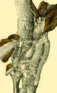
- Discussion:
- is apparent when with stress testing lateral tibial plateau rotates posteriorly in relation to the femur with lateral opening of joint;
- atraumatic type:
- presents as chronic laxity, often with a varus knee deformity;
- this ligamentous laxity often occurs without ACL or PCL laxity;
- external rotation of the lateral tibial plateau occurs around the intact PCL axis;
- in cases with significant varus deformity, a high tibial osteotomy may be the best treatment;
- ref: Proximal Tibial Opening Wedge Osteotomy as Initial Treatment for Chronic Posterolateral Corner Deficiency in the Varus Knee: a Prospective Clinical Study
- traumatic type:
- mechanism: knee hyperextension combined with a varus stress;
- posterolateraly directed blow to the anteromedial tibia with the knee in hyperextension is most common mechanism;
- most often associated w/ either ACL or PCL injury, and is commonly found in knee dislocations;
- it is important to distinguish this type of instability from one plane posterior instability;
- isolated PCL/ACL reconstruction will correct the one plane instability but will not correct rotatory instability (thus comprimising postoperative result);
- posterolateral injury components:
- disruption of popliteus tendon;
- arcuate ligament complex (partial or complete);
- LCL and lateral capsular ligament (distally based injury (from fibula) is most common);
- references:
- Biomechanical and Anatomical Assessment After Knee Hyperextension Injury
- Clinical Exam Findings:
- reference: A Clinically Relevant Assessment of Posterior Cruciate Ligament and Posterolateral Corner Injuries. Evaluation of Isolated and Combined Deficiency
- Radiographs:
- look for abnormal widening of the lateral joint space;
- avulsion of the Gerdy tubercle off of the tibia;
- Segond fracture (lateral capsular sign);
- avulsion of the lateral aspect of the capsule from the tibial plateau;
- also seen with ACL tears;
- consider arteriogram if dislocation is possible;
- MRI findings:
- bone contussion to medial femoral condyle;
- distal avulsion of LCL;
- fibular head fracture or osseous avulsion (arcuate ligament injury);
- reference: MR evaluation of the "arcuate" sign of posterolateral knee instability.
- Non Operative Treatment:
- grade-I and many moderate grade-II injuries can be treated nonoperatively;
- w/ grade II injuries, residual laxity may remain with non operative treatment;
- Surgical Treatment:
- preop planning:
- MRI allows assessement of posterolateral corner injury as well as ACL / PCL;
- MRI may help assess which structures of the posterolateral corner are injured (biceps, LCL, ect), and may help determine whether the injuries are mid-substance or whether they have been avulsed from the fibula or femur;
- in the acute phase of the injury, these structures may be anatomically repaired
- in chronic cases, extensive scarring makes definition of the individual structures and subsequent anatomic restoration impossible;
- prior to soft tissue reconstruction, any concomitant genu varum deformity (or varus stress during gait) should be corrected
beforehand with a high tibial osteotomy;
- ref: Proximal Tibial Opening Wedge Osteotomy as Initial Treatment for Chronic Posterolateral Corner Deficiency in the Varus Knee: a Prospective Clinical Study
- when reconstruction is planned consider whether a tibial based vs a fibular based reconstruction is appropriate;
- arthroscopic findings:
- expect to find an abnormal "drive through sign" in which there is more than 1 cm of lateral opening and exceptional posterior visualization of lateral meniscus;
- note risks of fluid extravasation w/ acute injuries due to capsular damage;
- this can be prevented by firmly wrapping the ankle and calf with Coband;
- ref: Arthroscopic evaluation of the lateral compartment of knees with grade III posterolateral knee complex injuries.
- exposure:
- need to identify the IT band, hamstrings, fibular head, peroneal nerve, and femoral attachment of the LCL;
- incision:
- straight lateral incision centered over the lateral joint line;
- proximally the subQ flaps are mobilized to allow identification of the anterior and posterior borders of the IT band;
- the anterior and posterior attachments of this band are freed to allow anterior and posteiror mobilization;
- peroneal nerve is identified posterior to biceps and is followed distally around fibular neck (look for evidence of nerve injury);
- ref: Displacement of the common peroneal nerve in posterolateral corner injuries of the knee.
- sequential assessment of injury:
- look for avulsion of IT band off of Gerdy's tubercle, peroneal nerve injury, biceps avulsion off of the fibular head, LCL injury (proximal or distal), and popliteus avulsion;
- repair will procede from the deepest structures to the most superficial structures;
- IT Band:
- note that the posterior 1/3 of the IT band attaches to the femoral epicondyle;
- if this attachement is deficient, it should be repaired to help restore lateral stability;
- ref: The anatomy of the iliopatellar band and iliotibial tract.
- biceps tendon:
- in cases of avulsion off of the fibula, reattachement may be completed with either bone anchors or w/ a suture pull thru technique;
- when the biceps remains intact, a distally based flap of a portion of the biceps may be used to recreate the anatomy of the LCL;
- with this technique, a tenodesis of the biceps femoris tendon to the lateral femoral epicondyle is performed;
- this serves to reconstruct the LCL ligament;
- LCL:
- w/ mild redundancy, consider imbrication to the reconstructed LCL;
- if ligament is present, consider advancement on its femoral attachment;
- when the biceps remains intact, a distally based flap of a portion of the biceps may be used to recreate the anatomy of the LCL;
- if ligament is absent, then consider reconstruction w/ Achilles allograft;
- allograft reconstruction:
- w/ chronic posterolateral injury, Achilles tendon allograft may be indicated;
- main goal is to create a checkrein to external rotation;
- at the level of Gerdy's tubercle, a bone tunnel is created in the posterolateral tibia, just medial to fibular head;
- attachment of the IT band to the intermuscular septum may have to be freed for optimal exposure;
- allograft bone plug (9 mm graft and tendon) is contoured to fit tunnel, and is secured w/ an interference screw;
- tendinous portion of the graft is then secured in the region of the popliteus insertion w/ a bone anchor;
- the anchor site should not allow more than 3 mm of motion w/ knee flexion and extension;
- with this technique, the strong stability provided by the allograft may help compensate for disruption of the arcuate complex;
- references:
- Reconstruction of the Lateral Collateral Ligament of the Knee with Patellar Tendon Allograft.
- The insertion geometry of the posterolateral corner of the knee.
- Relative role changing of lateral collateral ligament on the posterolateral rotatory instability according to the knee flexion angles: a biomechanical comparative study of role of lateral collateral ligament and popliteofibular ligament.
- popliteus
- resists excessive external rotation of the tibia during knee flexion from 20 to 130 deg;
- w/ avulsion attempt to reattach popileus to its femoral attachment (bone anchor) and to its fibular head attachment (pull through sutures);
- w/ severe injury, consider tenodesis of popliteus from its femoral attachement to posterolateral corner of tibia (this may be augmented w/ IT band fascia);
- in some cases, popliteus may be injured at its musculoskeletal junction, which is more difficult to repair;
- in cases in which fibular and tibial insertions of popliteus are torn, single split achilles-tendon allograft can be used;
- the bone graft is inserted in the lateral femoral condyle and the graft is splint longitudinally and is then anchored into the proximal parts of the tibia and fibula;
- references:
- The popliteus tendon and its fascicles at the popliteal hiatus: gross anatomy and functional arthroscopic evaluation with and without anterior cruciate ligament deficiency.
- The insertion geometry of the posterolateral corner of the knee.
- The role of the popliteofibular ligament and the tendon of popliteus in providing stability in the human knee.
- arcuate ligament complex / popliteal-fibular ligament: (see arcuate ligament);
- reconstruction/repair of this structure is necessary to avoid excessive tibial rotation, especially as the knee moves from extension to flexion;
- remember that biceps tendon, LCL, and arcuate ligament all insert on fibular styloid, and that if there is a fibular styloid avulsion, osseous reattachement will restore all three structures;
- generally this structure will be injured when significant posterolateral instability is present;
- if ligament has not been avulsed off of fibular styloid, then look for injury as ligament passes vertically to its femoral attachment;
- references:
- The insertion geometry of the posterolateral corner of the knee.
- The popliteofibular ligament: rediscovery of a key element in posterolateral instability.
- The Posterolateral Attachments of the Knee. A Qualitative and Quantitative Morphologic Analysis of the Fibular Collateral Ligament, Popliteus Tendon, Popliteofibular Ligament, and Lateral Gastrocnemius Tendon.
- The role of the popliteofibular ligament and the tendon of popliteus in providing stability in the human knee.
- Anatomic Posterolateral Knee Reconstructions Require a Popliteofibular Ligament Reconstruction Through a Tibial Tunnel
- lateral meniscus:
- lateral meniscus repair
Classification of knee ligament instabilities. Part II. The lateral compartment.
Acute posterolateral rotatory instability of the knee.
Chronic posterolateral rotatory instability of the knee.
Posterolateral instability of the knee.
Posterolateral instability of the knee.
Operative treatment of posterolateral instability of the knee.
The structure of the posterolateral aspect of the knee.
Treatment of acute and chronic combined anterior cruciate ligament and posterolateral knee injuries.
The posterolateral aspect of the knee. Anatomy and surgical approach.
The docking technique for posterolateral corner reconstruction.
Mechanical Properties of the Posterolateral Structures of the Knee.
The Posterolateral Corner of the Knee: Repair Versus Reconstruction.
An Analysis of the Causes of Failure in 57 Consecutive Posterolateral Operative Procedures.
Femoral Fixation Sites for Optimum Isometry of Posterolateral Reconstruction
Double-bundle PCL and Posterolateral Corner Reconstruction Components are Codominant.

