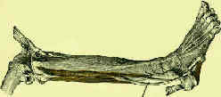
- Anatomy:
- lateral compartment
- plantar muscles of the foot:
- origin:
- lateral condyle of tibia, head and proximal 2/3 of lateral surface of fibula, intermuscular septa and adjacent fascia;
- course:
- peroneus longus courses posteiror to the brevis tendon, and then both tendons pass thru the common peroneal synovial sheath,
about 4 cm proximal to the lateral malleolus;
- synovial sheath passess through a fibro-osseous tunnel that is stabilized by the superior peroneal retinaculum and by the calcaneofibular
ligament;
- after the peroneus longus emerges from its sheath and passes inferior to the cuboid on its way to its insertion;
- os peroneum is an accessory bone which is located within the peroneus longus tendon in about 20% of feet;
- typically this is located plantar to the cuboid, near the calcaneo-cuboid articulation;
- this accessory ossicle may be associated with peroneus longus tenosynovitis;
- insertion:
- lateral margin of plantar surface of 1st cuniform and proximal end of 1st metatarsal.
- action:
- primary action is to plantar flex the first ray of the foot;
- plantar flexion and eversion of the foot at the ankle;
- primarily active during the stance phase of gait;
- gives lateral stability to the ankle;
- synergists: gastrocnemius, soleus;
- nerve supply:
- peroneal, S1 > L5, L4; (see innervation)
Pathologic Conditions:
- peroneal tendon subluxation:
- peroneal tendon disruption:
- persistent swelling along the peroneal tendon sheath is a reliable sign for peroneus brevis tendon tear;
- peroneal muscle spasm:
- may occur w/ tarsal coalition, but may also occur w/ rheumatoid arthritis, osteochondral frx, & infection in subtalar joint or
neoplasm (osteoid osteoma, fibrosarc)
- Charcot Marie Tooth:
- when there is loss of the tibialis anterior and peroneus brevis (which is common in CMT), the peroneus longus will cause flexion
of the first ray, which subsequently leads to a cavus foot deformity;
- polio:
- in polio syndrome, transfer of the peroneus longus in the presence of strong tibialis anterior results in a dorsal bunion as the
forefoot supinates;
- it must be combined with lateral transfer of the tibialis anterior to the base of the second metatarsal bone
Peroneal tendon injuries.


