

- Discussion:
- the Coventry type of HTO, creates an osteotomy above the tibial tubercle;
- in choosing a closing wedge osteotomy, the surgeon should be sure that the major deformity is on the tibial side (vs femoral side);
- the disadvantages of this type of osteotomy include possible non union, neurologic injury, and patellar baja;
- outcomes:
- in the study by Odenbring et al (1990), 75% of patients under the age of 50 w/ early medial DJD had at good result at 11 years
post surgery;
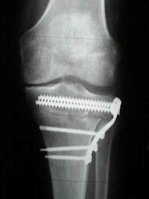
- in the report by Billings A, et al (1999), 64 valgus producing high tibial osteotomies were performed using a calibrated cutting
guide w/ plate fixation;
- 43 out of 64 knees had a good to excellent clinical result w/ knee score of 94 points at an average of 8.5 years follow up;
- using total knee arthroplasty as an end point, there was 85 % survival at 5 yrs and 53 % at 10 years;
- no patient had patella baja postoperatively (the authors fell that early ROM w/ CPM prevented baja);
- average initial postoperative correction (and standard deviation) for all knees was to 9.2 ± 3.69 degrees of valgus;
- 5 knees were corrected to less than 5 deg of valgus; 3 of them were treated with a subsequent arthroplasty (at twenty-four,
sixty-five, and sixty-six months);
- 13 knees had lost more than 2 deg of correction at the time of the latest follow-up;
- average initial postoperative correction for these knees was to 9.4 ± 4.12 deg (range, 4 to 17 degrees) of valgus;
- of knees that lost more than 2 degrees of correction, four subsequently had a total knee arthroplasty.
- references:
- Ten year results of tibial osteotomy for medial gonarthrosis: the influence of overcorrection.
- High tibial osteotomy with a calibrated osteotomy guide, rigid internal fixation, and early motion. Long-term follow-up.
- Revision after osteotomy for gonarthrosis: a 10-19 year follow up of 314 cases.
- Proximal tibial osteotomy in patients who are fifty years old or less. A long-term follow-up study.
- High tibial osteotomy. A prospective clinical and roentgenographic review.
- Cartilage regeneration after proximal tibial osteotomy for medial gonarthrosis. An arthroscopic, roentgenographic, and histologic study.
- Results of proximal tibial osteotomy. The effects of tibiofemoral angle, stance-phase flexion-extension, and medial-plateau force.
- Proximal osteotomy of the tibia for the treatment of genu recurvatum in adults.
- Factors influencing long-term results in high tibial osteotomy.
- Valgus high tibial osteotomy. A long-term follow-up study.
- High tibial osteotomy.
- A ten- to 15-year follow-up observation of high tibial osteotomy in medial compartment osteoarthrosis.
- Proximal tibial osteotomy. A critical long-term study of eighty-seven cases.
- Proximal tibial osteotomy. Factors that influence the duration of satisfactory function.
- High tibial osteotomy. A prospective clinical and roentgenographic review.
- Proximal tibial osteotomy for osteoarthritis with varus deformity. A ten to thirteen-year follow-up study.
- Valgus high tibial osteotomy. A long-term follow-up study.
- Tibial osteotomy for osteoarthritis of the knee. A five to ten-year follow-up study.
- Tibial Osteotomy for the Treatment of Varus Gonarthrosis. Survival and failure analysis to twenty-two years.
- General Concept:
- incision:
- inverted L shaped incision;
- transverse limb:
- is made at the level of the joint line (or just below it);
- posteriorly the incision extends just past the fibular head;
- anteriorly the incision extends to the patellar tendon;
- verticle limb:
- extends inferiorly along the lateral crest of the tibia for 10 cm;
- superiorly, taken care not to injure the patellar tendon;
- incise thru the patellar paratenon and bluntly spread beneath the tendon;
- during the osteotomy cuts, a spade retractor can be placed just underneath the patellar tendon for protection;
- incision is carried down to periosteum, and anterior compartment musculature is elevated off the cortical surface;
- proximally the incision follows the posterior aspect of biceps tendon inorder to expose the peroneal nerve;
- distally the incision courses obliquely and horizontally to a point just distal to the tibial tubercle;
- a Chandler "spade" retractor is placed just anterior to the peroneal nerve and a second Chandler is placed just posterior to the
patellar tendon;
- proximal cut is made 1.5 cm below and horizontal to joint line;
- proximal cut should avoid transecting the posterior tibial cortex, since it is desirable to have the posterior cortex of the
proximal fragment overlap the proximal cortex of the distal fragment;
- distal cut is usually made 7 mm to 1 cm below the proximal cut;
- management of fibula:
- Method of Fixation: (controversial);
- plate fixation: (procedure of choice)
- offers early mobilization of knee joint & has shortened rehab time and decreased the incidence
of patella infera;
- advantage: does not require immobilization of the knee;
- disadvantage: does not allow full wt bearing;
- staple fixation:
- Post Operative Care:
- importance of early ROM;
- w/ closing wedge osteotomy, distance between tubercle and joint line is decreased, decreasing tension on patellar ligament;
- patellar ligament, fat pad, and retinaculum may contract and scar causing patella baja;
- Billings A, et al. (2000), no patient had patella baja postoperatively (the authors felt that early ROM w/ CPM prevented baja);
- neurologic exam:
- depending on whether or not the peroneal nerve is decompressed and depending on how the fibula is managed (fibular
osteotomy vs tib-fib capsular transection), all patients will require an immediate postoperative peroneal nerve examination
as well as an examination the next day;
- a "delayed peroneal nerve palsy" may have two causes:
- in some patients, dense fibrous tissue will firmly anchor the peroneal nerve to the neck of the fibula;
- in this situation, postoperative edema in this region may cause nerve compression;
- alternatively, a "delayed peroneal nerve palsy" may represent an evolving comartment syndrome (which can be masked
if an epidural is used for postoperative anesthesia);
- ref: HTO with a calibrated osteotomy guide, rigid internal fixation, and early motion. Long-term follow-up.
- Controversies:
- management of varus laxity following HTO:
- this may occur due to relative shortening of the femoral to fibular head distance, causing laxity of the LCL;
- patients w/ a prominent varus thrust may be especially at risk for postop laxity;
- when recognized intraoperatively, consider posterolateral advancement;
- w/ excessive external rotation, then consider advancement of the popliteus;
- w/ excessive varus laxity, consider advancement of the LCL;
- both the popliteus and LCL may be advanced together using a common wafer of bone;
- references:
- Recurrent Varus Angulation After High Tibial Osteotomy: An Anatomic Analysis.