

- Discussion:
- surgical indications include diabetic neuropathy, osteoarthritis, posttraumatic injury, talar AVN, and RA involving
ankle and subtalar joints;
- position for pantalar arthrodesis:
- 0-5 deg of calcaneus (not equinus) and 5 deg of valgus;
- in the report by Chou, et al, the authors studied 55 patients (56 ankles) who underwent simultaneous tibiotalocalcaneal
arthrodesis with severe disease involving the ankle and subtalar joints;
- average time of follow-up was 26 months after the operation;
- fusion was achieved in 48 ankles, with an average time of fusion of 19 weeks;
- 48 of the 55 patients were satisfied with the procedure;
- average leg length discrepancy was 1.4 cm;
- average amount of dorsiflexion was 2 degrees and plantar flexion was 5 degrees;
- 42 patients complained of post op pain, 40 patients required shoe modification or an orthotic device, and 34 patients had a limp;
- most common complications were nonunion (8 ankles) and wound infection (6 ankles);
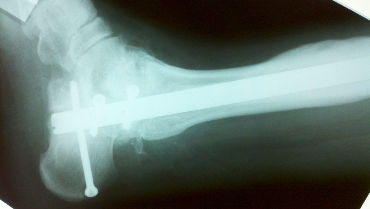
- Tibiotalocalcaneal arthrodesis.
- Technique Considerations:
- need to keep foot plantigrade:
- if foot cannot be positioned plantigrade, then consider need for partial or complete talectomy;
- medial approach to ankle and subtalar joint:
- ensures that there will be no neurovascular injury;
- tarsal tunnel decompression sometimes affords improved sensation to the foot;
- retrograde nails:
- which are used for pantalar fusion should have interlocking nails in the saggital plane inorder to counteract the muscular forces generated in gait;
- using the standard technique, the lateral plantar nerve and artery are at risk for injury, not to mention muscle and tendon injury (esp to the FHL tendon);
- in the study by McGarvey WC, et al (1998), medial malleolar osteotomy and medial translation of the talus, reduces the risk of N/V
injury, FHL injury, and increases the strength of fixation;
- Tibiotalocalcaneal arthrodesis: Anatomic and technical considerations.
- distal interlocking screws;
- in most systems one of the interlocking screws will traverse in the AP direction - consider making this screw extra-long so that
it engages the midfoot for additional fixation;
- Complications:
- Limb salvage: the infected retrograde tibiotalocalcaneal intramedullary nail.
Minimally invasive ankle arthrodesis with a retrograde locking nail after failed fusion
Tibiotalocalcaneal arthrodesis for arthritis and deformity of the hind part of the foot.
Tibiotalocalcaneal Arthrodesis Using a Reamed Retrograde Locking Nail
Ankle arthrodesis with a retrograde femoral nail for Charcot ankle arthropathy.
Charcot ankle fusion with a retrograde locked intramedullary nail
Tibiotalocalcaneal arthrodesis using a dynamically locked retrograde intramedullary nail.
Ankle fractures in diabetic neuropathic arthropathy: can tibiotalar arthrodesis salvage the limb?
.............................................................................................................................................................................................................................................................................................................................
Original Text by Clifford R. Wheeless, III, MD.
Last updated by Clifford R. Wheeless, III, MD on Thursday, July 9, 2015 4:23 pm