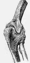
- See:


- Posterior Approach:
- Lateral Approach
- Position:
- lateral w/ the arm abducted 90 deg;
- force of gravity maintains the correct position;
- sterile tourniquet is applied;
- for simple fractures: supine with the arm laid across the chest;
- the position must allow flexion and extension;
- Incision:
- incision is started posteriorly above elbow, and continues distally, curving either ulnarly or radially to avoid point of
olecranon, & then continue still distally towards the posterior border of ulna to a point 3-4 cm distal to frx;
- ulnarly based incision is a better choice since it affords better exposure of ulnar nerve which may need to be transposed
anteriorly at end of case;
- a radial incision will require considerable underming;
- olecranon bursa is incised and no effort is made to protect it;
- aconeus is elevated on the lateral aspect of the ulna;
- idenitify and preserve the radial and ulnar collateral ligaments;
- Frx Visualization and Reduction:
- performed w/ elbow in extension which relaxes pull of triceps muscle;
- frx site is visualized by elevating periosteum proximally w/ proximal fragment;
- articular surface is visualized laterally and medially by elevating the triceps retinaculum and periosteum;
- lateral exposure requires detachment of aconeus muscle from radial side of the ulna;
- medial exposure risks ulnar nerve injury;
- frx is reduced and held w/ one or two towel clips or K wires;
- begin by drilling a superficial hole in the distal fragment to allow introduction of the tip of the pointed reduction forceps;
- other tip of forceps catches proximal fragment & reduces fracture;
- Choice of Fixation:
- Tension Band Wiring:
- Plate Fixation

