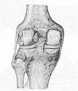
- Discussion:
- the ligaments of Humphrey and Wrisberg are meniscofemoral ligaments which run from the posterior horn of the
lateral meniscus to the lateral aspect of the medial femoral condyle;
- these ligaments are named based on their location in relation to the PCL;
- the anterior meniscofemoral ligament is known as the ligament of Humphrey where as the posterior meniscofemoral
ligament is known as the ligament of Wrisberg;
- in about 70 % of knees, there is either anterior meniscofemoral ligament of Humphrey or posterior meniscofemoral
ligament of Wrisberg;
- latter is more common and is characterized by femoral origin merging w/ that of posterior cruciate ligament;
- in 6% of knees, both ligaments will be present;
- these meniscofemoral ligaments may play minor role as secondary restraints to posterior tibial translation after complete
transection of the posterior cruciate ligament;
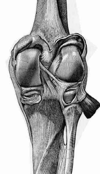
- Humphrey ligament: (anterior meniscofemoral ligament);
- is less than 1/3 the diameter of the PCL;
- arises from the posterior horn of the lateral meniscus, runs anterior to the to the PCL and inserts at the distal edge of the femoral PCL
attachment;
- may be confused for the PCL during arthroscopy;
- in this situation, tug on the ligament while observing for motion of the lateral meniscus;
- Wrisberg's ligament: (posterior meniscofemoral ligament);
- usually larger than ligament of Humphrey (upto 1/2 the diameter of the PCL diameter);
- extends from the posterior horn of lateral meniscus to medial femoral condyle
Anatomical and biomechanical characteristics of human meniscofemoral ligaments.
The menisco-femoral ligaments.
The meniscofemoral ligaments of the knee.
Meniscofemoral ligaments revisited. Anatomical study, age correlation and clinical implications