

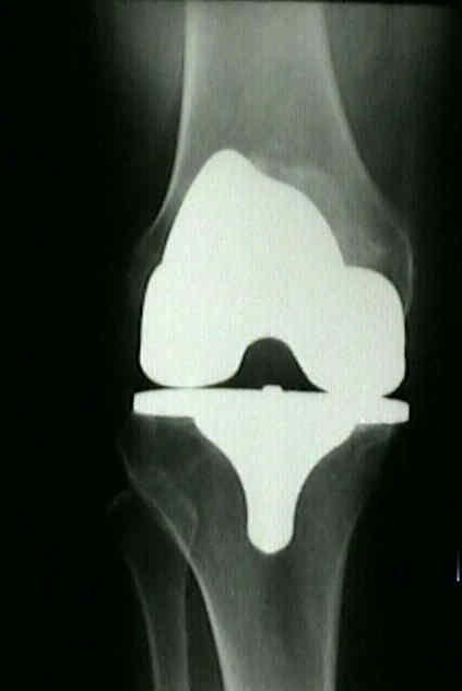
- See: PreOp Planning:
- Discussion:
- a candidate for HTO should have relatively normal lateral and patellofemoral compartments;
- degenerative changes confined to medial compartment should be apparent in x-rays, although minor arthritic changes in lateral
compartment or patellofemoral joint are not absolute contraindications
- HTO is contraindicated w/ lateral compartment or patellofemoral symptoms along with associated radiographic changes;
- Radiographs: 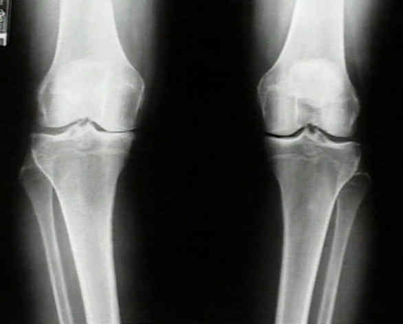
- full-length, wt bearing x-rays are used to determine the desired amount of correction;
- to unload the arthritic medial compartment, a relative valgus knee alignment is created;
- radiographic methods for planning the osteotomy:
- mechanical axis method:
- requires long cassett;
- the mechanical axis is represented by the angle formed by the line joining the femoral head to the center
of the the intercondylar notch and the line joining the interspinous region to the center of the ankle;
- normally this line should be superimposed over the wt bearing axis;
- if neutral alignment is considered noral, 2-3 deg overcorrection would be indicated for the patient with medial joint arthrosis;
- anatomic axis method:
- formed by the intersection of lines formed by the axis of the femur to the intercondylar notch and the line formed by the
interspinous region to the center of the ankle;
- goal is to achieve 8 deg of valgus;
- wt bearing axis is represented by the line drawn thru center of femoral head to the center of the ankle;
- normally it should pass just medial to the center of the knee joint;
- if neutral alignment is considered normal, 2-3 overcorrection would be indicated for the patient with medial joint arthrosis;
- supine over correction method:
- motivation for this technique is based on the premise that patients whose varus deformity is based in part on ligamentous laxity
need less correction than patients w/o laxity;
- assumptions: the mechanical axis value just prior to heel strike is similar to the supine mechanical axis measurement;
- measure the supine mechanical axis and add 10 deg of correction (supine overcorrection);
- Anatomy:
- varus or valgus angulation of arthritic knee usually results from 3 components:
- geometric alignment of femur & tibia;
- narrowing or loss of bone & cartilage in one compartment;
- widening of other compartment's joint space because of slack ligamentous and soft-tissue structures;
- detected by measuring difference between involved knee and the opposite, uninvolved knee.
- each millimeter of excessive joint space separation causes an apparent 1 deg of angular deformity on weightbearing x-ray;
- if widening is not taken into consideration malalignment may be overcorrected;
- Technical Considerations:
- its essential to have good quality radiographs both supine and standing;
- ensure that radiographs are taken w/ the patella facing forward (rather than the patient's feet);
- note the difference between wt bearing and non wt bearing radiographs
Axial parameters affecting lower limb alignment after high tibial osteotomy.