

- Septic Knee:
- Synovium of the Knee:
- Anatomy:
- four bursae around knee are susecptible to and inflammatory response from direct or indirect trauma;
- prepatellar bursae is most commonly affected area (housemaids knee);
- may show significant degree of swelling;
- two bursae are infrequently affected;
- infrapatellar and deep patellar bursae;
- when dx is in must also consider fat pad impingment syndrome versus bursitis;
- fourth bursa:
- deep to pes arserinus insertion;
- rarely affected w/ bursitis (dx of exclusion)
- first r/o chondral frx, meniscal tear, or osteonecrosis;
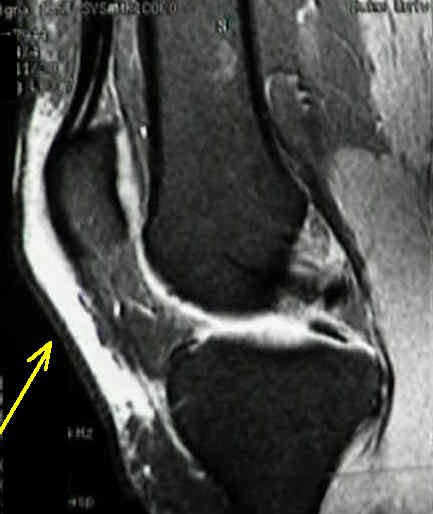
- Infrapatellar bursitis:
- small deep subpatellar or infrapatellar bursa is located between tuberosity of tibia & patellar tendon and is separated from synovium of
the knee by a pad of fat;
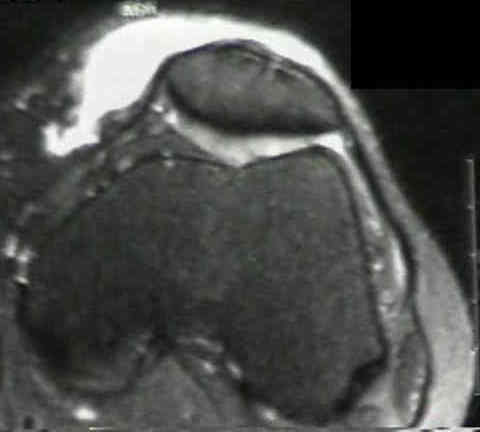
- Prepatellar Bursitis:
- traumatic prepatellar bursitis may be caused by acute injury such as fall directly on the patella or by recurrent minor injuries, such as
those that produce "housemaid's" knee;
- pyogenic prepatellar bursitis is common, especially in children;
- when bursae is large, swelling may be so pronounced that dx of pyogenic arthritis of knee joint may be mistakenly made;
- this mistake must be avoided because if the knee joint is opened pyogenic arthritis will develop;
- on other hand, if correct dx is made & bursa is drained properly, pyogenic arthritis is prevented;
- Management of Bursitis:
- aspiration and injection of an appropriate drug;
- traumatic bursitis will often respond favorably to aspiration & injection of an appropriate steroid preparation;
- incision and drainage when an acute suppurative bursitis fails to respond to non surgical treatment;
- excision of chronically infected and thickened bursae
- removal of underlying bony promineces;
- Technique of Drainage:
- approach of bursa thru two longitudinal incisions, one medial and one lateral, or thru a single transverse incision;
- open bursa, evacuate its contents, and pack it loosely w/ petrolatum gauze or close it loosely over a drain as seems appropriate;
- compression dressing should be applied after aspiration;
- After Treatment:
- because cellulitis is always present, the extremity is immobilized in posterior splint, and appropriate antibiotics are given;
- if gauze has been used to pack bursa, it is changed at least qod;
- even w/ good drainage, sinuses often persist on one or both sides of joint;
- joint must not be invaded since bursa does not communicate w/ it;
- pt should be informed when first seen that complete excision of of bursa may be necessary if healing fails to occur after simple
drainage;
- when walls of bursa are thickened from chronic inflammation, resecting entire bursa is usally easy, but when lesion is acute &
effusion is serous, excising the bursa completely may be impossible, yet enough may be excised to relieve symptoms;
- occassionally fibrosis or synovial thickening w/ painful nodules requires excision of the bursae
The skin incision in the excision of the prepatellar bursa
Prepatellar and olecranon bursitis: literature review and development of a treatment algorithm