

- Radiographic Findings:
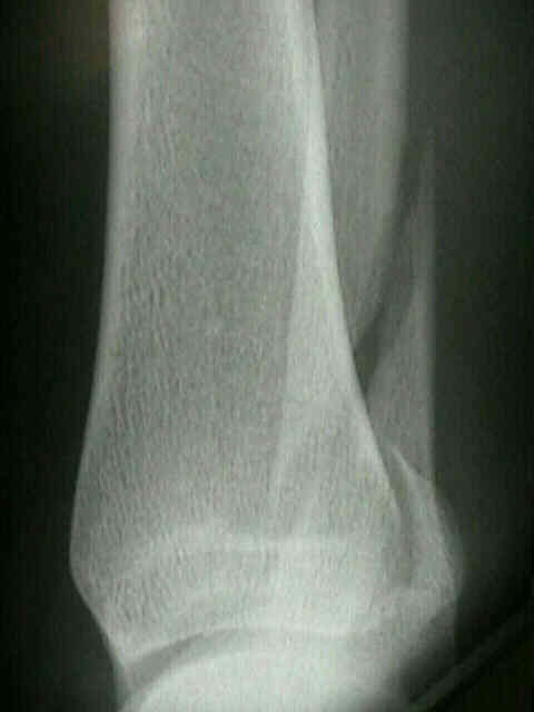
- ankle frx:
- fibula overlaps posterior aspect of tibia, but posterior tubercle of tibia can still be seen;
- posterior subluxation of talus in mortise may be assoc w/ posterior lip frx or posterior talofibular ligament tear;
- stress lateral view:
- correlates well w/ anatomic diastasis;
- is more sensitive than mortise view in determining syndesmotic injury;
- w/ a 7 mm anatomic diastatis, lateral malleolus may posteriorly displace 4-5 mm, where as the Mortise view shows only 1-2 mm of displacement;
- ref: The tibiofibular syndesmosis. Evaluation of the ligamentous structures, methods of fixation, and radiographic assessment.
- talus
- talar neck frx:
- lateral talar process frx
- os trigonum:
- is relatively frequently seen accessory bone of foot found just posterior to lateral tubercle of posterior process;
- anterior impingment syndrome
- anterior widening:
- dome of talus is centered under the tibia and congruous w/ distal tibial articular surface;
- asymmetry of articular space, esp anterior widening, suggests instability