

- Discussion:
- flex the knee to 90 deg and release the anterior attachment of medial joint capsule from the tibia;
- this should provide wide exposure of the interior of the knee;
- at this point it should be possible to sublux tibia forward for complete visualization of the plateau surfaces;
- trim osteophytes from articular margin of tibia, femur, & patella with a rongeur or an osteotome;
- cushing rongeur is used to remove osteophytes in medial & lateral aspects of the femoral condyles as well as the intercondylar space;
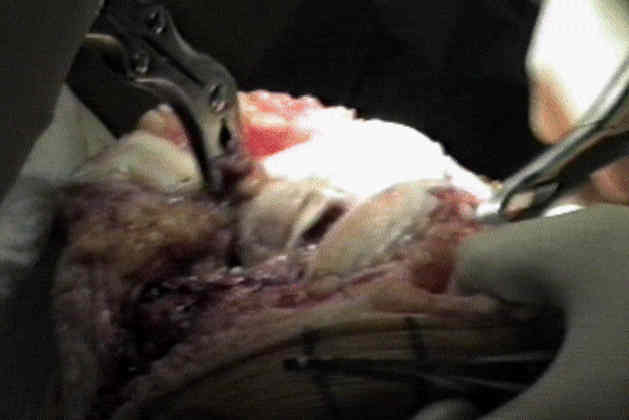
- take care to remove any osteophytes from intercondylar notch of femur, particularly if prosthesis is designed to retain posterior cruciate;
- now assess the overall alignment of the leg;
- if full extension is possible & gentle Varus or Valgus stress will align knee, significant release will probably not be necessary;
- anterior halves of menisci are easily excised, leaving posterior halves to be excised after resection of bone from femur, at which time they are readily accessible;
- if synovial lining is hyperplastic, excise portions that interfere w/ performing the arthroplasty (complete synovectomy not necessary);
- 90 deg Homan retractor is positioned between the everted patella & distolateral femur, exposing the lateral patellofemoral ligament, which is incised with electrocautery;
- retractor is repositioned in the interval between iliotibial tract & tibial attachment of the capsule;
- capsule is dissected free from the infrapatellar fat pad;
- lateral inferior geniculate artery is ligated;
- insertion of the iliotibial tract is identified, and the capsule dissected from the lateral tibial condyle;
- remove all osteophytes, then clear out posterior & medial osteophytes, & then adress those along ridges & rims & around PCL