- See:
- soft tissue reconstruction of the leg
- fracture blister management
- Discussion:
- based on the injury to the soft tissue and whether the injury resulted from high or low injury trauma;
- type I & II frx
- closed reduction, cast or splint application, and elevation;
- immediate ORIF can be considered if there is not excessive swelling or excessive soft tissue disruption;
- type III B and C frx
- operative timing and strategy:
- goal is to keep talus centered under the tibia and fracture out to length while soft tissue heal over 7 to 21 days;
- early medial incisions in the face of tense soft tissue swelling, risks wound infection and slough;
- the greatest risks seems to occur from ORIF of the medial column within 5 days of injury;
- Sirkin M, et al (1999):
- immediate ex fix & ORIF of fibula, & formal ORIF of tibial articular surface performed on delayed basis
(avg delay 12-13 days);
- no patient that presented with a closed injury developed a full thickness skin necrosis and none required
secondary soft tissue coverage;
- authors feel that historically high rate of infection and skin necrosis following ORIF of these injuries is most
related to operative timing;
- Patterson MJ and Cole JD (1999), all patients underwent a two staged technique for treatment of complex pilon frx;
- initially all patients underwent immediate fibular fixation and placement of a medial fixator;
- Blauth M, et al.:
- authors recommend a 2-step procedure for the treatment of severe tibial pilon fractures with extensive soft
tissue damage;
- first stage:
- includes primary reduction and internal fixation of the articular surface is performed using stab incisions,
screws, and K-wires;
- temporary external fixation is applied across the ankle joint;
- second stage:
- requires recovery of the soft tissues;
- involves internal fixation with a medial plate using a reduced invasive technique;
- references:
- A staged protocol for soft tissue management in the treatment of complex pilon fractures
- Two-staged delayed open reduction and internal fixation of severe pilon fractures.
- Surgical Options for the Treatment of Severe Tibial Pilon Fractures: a Study of Three Techniques
- calcaneal pin:
- calcaneal pin w/ 15-20 pounds of traction with the leg elevated on a Bohler frame allows alignment of frx fragments and
brings frx out to length while soft tissues are monitored;
- external fixation:
- external fixation may substitute for calcaneal pin traction, and allows the frx to be brought out to length;
- may consist of a simple tibial fixator that includes the foot, or may consist of a more elaborate circular wire fixator;
- it is controversial whether or not to fix the fibula in these fractures, and it is further controversial as to whether to perform
ORIF with the initial fixator placement or to perform it on a delayed basis;
- note it is important to apply the fixator as medially as possible inorder to avoid the standard medial incision site (should it
be necessary for articular restoration);
- references:
- External fixation of severely comminuted and open tibial pilon fractures.
- Definitive plates overlapping provisional external fixator pin sites: is the infection risk increased?
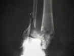
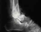
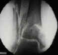
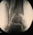
- case example: 40-year-old female w/ grade 3b open pilon frx (seen on left), who was treated w/ a hybrid fixator which
brought the frx out to length);
Does a staged posterior approach have a negative effect on OTA- 43C fracture outcomes?
Open reduction and internal fixation of tibial pilon fractures.
The management of the soft tissues in pilon fractures.
The results of early primary open reduction and internal fixation for treatment of OTA 43.C-type tibial pilon fractures: a cohort study.
Temporary bridging external fixation in distal tibial fracture.
Tibial pilon fractures: a review of incidence, diagnosis, treatment, and complications.
Complications and early results after operative fixation of 68 pilon fractures of the distal tibia.
Distal tibial fractures and pilon fractures.
[One-stage management of open distal tibial Pilon fractures].
Early limited internal fixation of diaphyseal extensions in select pilon fractures: upgrading AO/OTA type C fractures to AO/OTA type B.


