

 - See: Bone Tumor Menu
 - Discussion:
- rare tumor of unknown origin;
- occurs primarily in young males between 10 - 30 years of age (most common after skeletal maturity);
- pts present w/ firm, slowly enlarging mass that produces minimal disability;
- on other occassions the presenting features will be pain, swelling or pathologic fracture;
- sites of involvement:
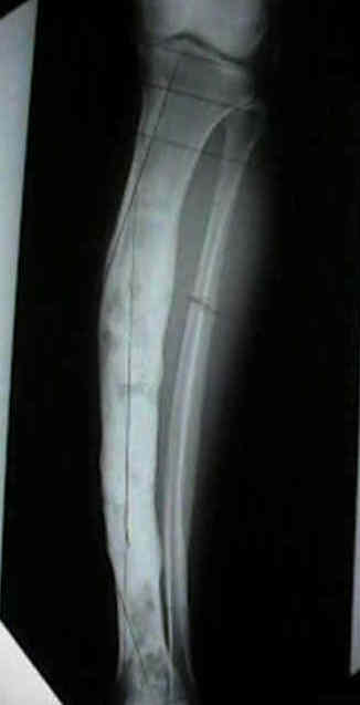
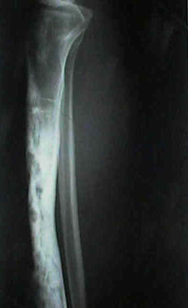
- mandible or tibial diaphysis is involved in 90% of pts;
- occassionally tumor arises in forearm, hands, or feet;
- histology:
- multiple biopsies may be required to have an representative sample;
- look for a biphasic pattern of glandular epithelial cells which are surrounded by fibrous spindle cells;
 - epithelial cells may be grouped in nests and stain positive for keratin;
- diff dx:
- chondromyxoid fibroma
- nonossifying fibroma
- fibrous dysplasia
- osteofibrous dysplasia:
- occurs most often in skeletally immature patients;
- most often involves the anterior cortex of the tibial diaphysis;
- minor anterior bowing of the tibia is frequently seen
- extensive histologic evaluation is necessary to distinguish lesions in the spectrum between osteofibrous dysplasia and adamantinoma;
- in some patients, this lesion will progress to full blown adamantinoma;
- there is some indication that curretage or marginal excision should not be undertaken because of high local recurrance rate;
- references:
- Osteofibrous dysplasia (ossifying fibroma of long bones): a report of four cases and review of the literature.
- Osteofibrous dysplasia of the tibia and fibula.
- Treatment:
- because these tumors are slow growing and have a limited potential for local recurrence or metastasis;
- marginal resection: delayed local recurrence can be expected in 32%;
- en bloc resection: local recurrence should not occur;
- mean survival w/ metastatic disease: 12 years;
- in report by Qureshi AA, et al (2000), authors examined the role of en bloc excision on 70 patients was examined;
- limb salvage was attempted in 91 percent (sixty-four) of the seventy patients, and the final rate of limb preservation was 84 % (59 of 70);
- wide operative margins were obtained in 92% (58) of 63 patients;
- intercalary allograft was used to reconstruct the segmental bone defect in 51% (36) of the 70 patients;
- reconstruction-related complications occurred in 48% (30) of 62 patients;
- nonunion and fracture were the most common complications, occurring in 24% (15) and 23% (14) of 62 patients, respectively.
- Kaplan-Meier analysis demonstrated a rate of local recurrence of 18.6 percent at ten years.
- XRT: adamantinoma is highly radioresistant
- chemotherapy has not been shown to be effective;
- Current trends in the management of adamantinoma of long bones. An international study.
Ten Most Common Bone and Joint Tumors--General Orthopaedics: Adamantinoma of the Appendicular Skeleton--Updated.
The treatment of adamantinoma of the tibia by wide resection and allograft bone transplantation.
Adamantinoma of the tibia masked by fibrous dysplasia. Report of three cases.
Adamantinoma of long bones. A clinicopathological study of fourteen cases with vascular origin suggested.
Adamantinoma of the long bones. A clinicopathological study of thirty-two patients with emphasis on histological subtype, precursor lesion, and biological behavior.