- Discussion:
- plain films may not reveal posterior arch frx, frx and subluxations of articular facets, frx of C1 and
C2 (among most commonly missed fractures on plain films), and interspinal bony fragments;
- plain films may not pick up the coronally oriented vertebral body frx resulting in large separate anterior tear drop fragment;
- to evaluate mid sagittal fracture thru the posterior vertebral wall;
- finally CT is best method for determing encroachment into bony canal;
- indications:
- indicated for unconscious patient w/ suspicious or inadequate cervical radiographs;
- CT is indicated w/ all cervical fractures or suspected fractues on initial plane films;
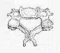
- delineating injuries to the atlantoaxial complex, esp rotatory subluxation and C-1 ring fractures;
- used to examine Jefferson Fx, rotary dislocation, burst fractures, cervicothoracic level injuries;
- Technique:
- request thin sections (1.5 - 3 mm thickness), either continguous or overlapped, with reformations in the saggital, coronal, and paraoblique planes (thru the neural foremen) when appropriate;
- images should be photographed twice, to optimize both soft tissue (intraspinal) and bone detail;
- CT begins one level above the frx and continue one level below frx;
- CT scan includes five-millimeter sections in a spiral format;
- use non-spiral format to minimize image-averaging artifact;
- Disadvantages of CT:
- CT scans are much less sensitive for detecting frx in transverse plane such as articular process fractures;
- oblique views are more sensitive than reformatted, overlapping, transverse CT cuts for detecting certain articular process injuries, and certainly are more readily available;
- risks of cancer:
- ref: Computed Tomography — An Increasing Source of Radiation Exposure
- Tomography
- for visualization of lateral masses using either linear or nonlinear motion patterns;
- tomography is helpful in defining extent of facet frx or subluxation;
- most units have vertical beam, requiring that pt be positioned lying on side to obtain lateral views;
- this can be awkward or dangerous w/ acute spine injury;
- tomography is particularly helpful in determining the anatomic level of a dens fracture
References
The role and limitations of computed tomographic scanning in the evaluation of cervical trauma.
Unsuspected upper cervical spine fractures associated with significant head trauma: role of CT.


