- Pathoanatomy:
- osteophytosis occurs as result of breakdown in the out annular fibers of annulus fibrosis;
- disk material stretching & displacing these fibers, causing stress at ligamentous attachments leading to formation of osteophytes;
- osteophytes collects initially extend horizontally;
- later on osteophytes extend vertically from edges of vertebra, sometimes bridging disk space;
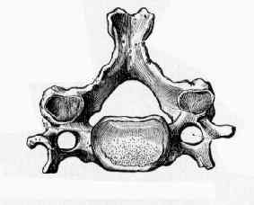
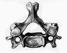
- involves the disc, two facet joints & two false uncovertebral joints (Lushka);
- cervical cord becomes impinged when diameter of canal (normally about 17 mm) is reduced to less than 13 mm;
- hyperextension:
- cord increases in diameter and it & roots are pinched between discs and adjacent spondylitic bars anteriorly, and hypertrophic
facets and infolded ligamentum flavum posteriorly;
- hyperflexion:
- cord narrows and the neural structures are tethered anteriorly across discs or spondylitic bars;
- radiculopathy:
- spondylotic changes in the foramina primarily from chondro-osseous spurs of the joints of Luschka may restrict motion and may
lead to nerve root compression;
- soft disc herniation:
- is usually posterolateral, between posterior edge of uncinate process & lateral edge of posterior longitudinal ligament, resulting in
acute radiculopathy;
- myelopathy:
- central herniation;
- spondylotic bars with a congenitally narrow canal;
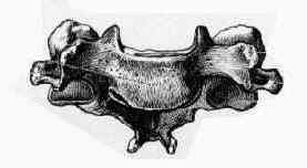
- Apophyseal Joints:
- show early irregularity and blurring of the joint surfaces;
- joint space narrowing and eventual spurring and sclerosis;
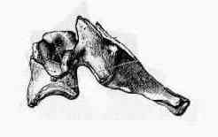
- lateral view & oblique view:
- allows evaluation of facet joints;
- determine if osteophytes of apophyseal joints project medially into foramina canal;
- specifically, osteophytes arising from the ventral portion of superior articular process may cause symptomatic foraminal narrowing;
- rarely osteophytes may also project anteriorly and impinge upon vertebral artery, resulting in arterial insufficiency;
- loss of disk height leads to reduced neuroforaminal volume, rendering root more susceptible to compression;
- Joints of Luschka:
- joints give rise to bony spurs or ridges -osteophytes- as can main fascet joints & edges of vertebral bodies adjacent to intervertebral disc;
- this is symphysis type of articulation between vertebral bodies;
- exiting nerve root on each side travels between these joints, & can be compressed by osteophytes extending into intervertebral
foramen from any or all three of sources mentioned