- See:
- Maisonneuve Fracture
- Surgical Approaches for Lateral Malleolar Fracture:
- Anterolateral Approach
- Posterolateral Approach
- Weber A Lateral Malleolar Frx
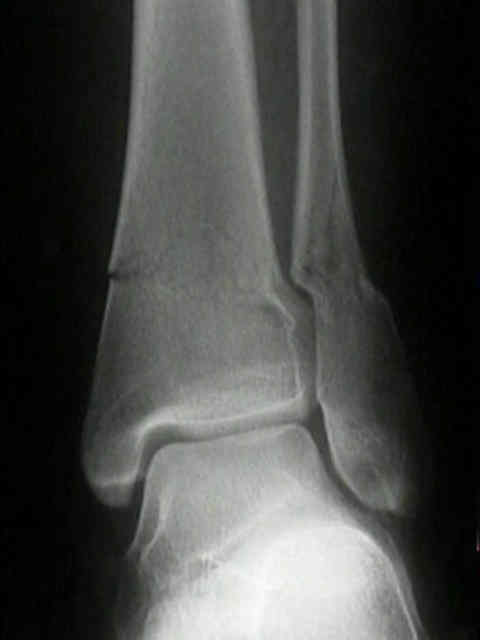
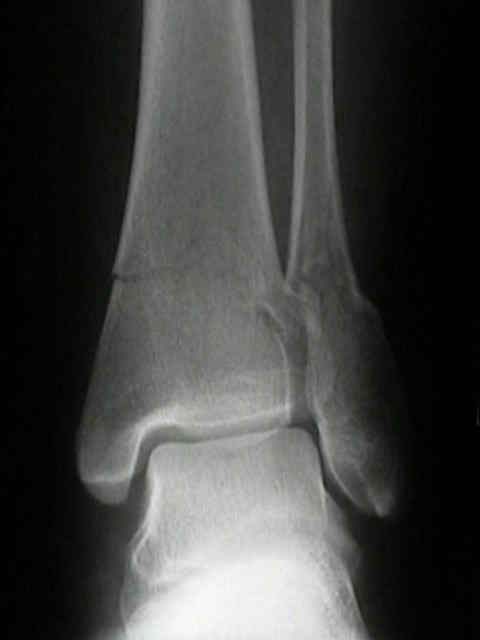
- Weber B Lateral Malleolar Frx
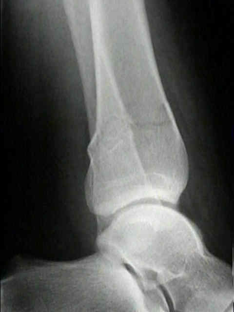
- Weber C: Lateral Malleolus
- Discussion:
- malposition of lateral malleolus w/ tilting or shortening, will lead to distorted talotibial wt bearing and lead to rapid degenerative arthritis;
- if lateral displacement of talus is not corrected, granulation tissue grows into medial joint space & is converted to fibrous tissue, which prevents late closed reduction of the displacement;
- for ankle to function satsifactorily, fibula must be:
- normal in length
- correctly positioned in the groove of the tibia
- effectively anchored to tibia through syndesmosis;
- anatomic restoration of distal tibiofibular syndesmosis is needed;
- fibula is primary resistance to external rotation forces on ankle;
- tendons of the long calf muscle controlling ankle joint tend to displace the talus laterally;
- because the lateral collateral ligaments secure the fibula to the talus, the talus follows the fibula after fracture;
- if lateral talar shift is greater than lateral displacement of fibula, there may be an injury the sysdesmosis;
- frx of fibular malleolus, even w/ other structures intact, results in 25 deg of ext rot instability & 1-2 mm of lateral talar displacement;
- 1 mm of talar shift decreases the tibiotalar contact by 50% (in a cadaver model);
- complications:
- most common complication after fracture of the fibula is malunion;
- this is usually in the form of posterior and external rotation displacement of the maleolus and fibular shortening;
- this disturbs the ankle by allowing external rotation of the talus and lateral talar shift;
- Operative Treatment:
- indications:
- w/ medial tenderness, > 5 mm of medial clear space (on static or stress views) diagnosis of injury of the deltoid ligament can be made;
- treat as bimalleolar frxs, w/ ORIF of lateral malleolus;
- surgical approaches:
- reduction: weber B fractures:
- oblique fractures:
- w/ oblique or spiral frx that is not comminuted, fix it with one or two lag screw inserted from anterior to posterior to achieve intrafragmentary compression;
- screws should engage the posterior cortex but should not protrude far enough posteriorly to encroach on the peroneal tendon sheaths;
- plate position
The key role of the lateral malleolus in displaced fractures of the ankle.
Reconstruction of malunited fractures of the lateral malleolus.
The Dorsal Antiglide Plate in the Treatment of Danis-Weber Type-B Fractures of the Distal Fibula.