

- occurrence of syndesmosis injury with Weber B / SER fractures
- references:
- Intraoperative diagnosis of syndesmosis injuries in external rotation ankle fractures.
- Syndesmotic instability in Weber B ankle fractures: a clinical evaluation.
- Intraop Assessment of Stability of Distal Tibiofibular Joint in SER Injuries: Sensitivity, Specificity, and Reliability of 2 Clinical Tests
- Stability-based classification for ankle fracture management and the syndesmosis injury in ankle fractures due to a SER mechanism of injury.
- Syndesmotic fixation in supination-external rotation ankle fractures: a prospective randomized study
- Exam:
- direct palpation: palpate for tenderness over the syndesmosis;
- squeeze test: at the mid-calf, squeeze the fibula against the tibia and look for symptoms at the syndesmosis;
- external rotation test: forced external rotation may cause pain at the syndesmosis;
- PreOperative Radiographs:
- fibular fractures that begin proximal to tibial plafond are assumed to have some degree of injury of the syndesmosis;
- dorsiflexion and external rotation will accentuate syndesmotic widening;
- mortise view:
- tibiofibular overlap: should normally be more than 1 mm;
- lateral talar shift:
- tibio-fibular clear space:
- tibiofibular clear space> (interosseous clear space) is carilaginous space bewtween lateral border of posterior tibia (incisura fibularis) &
medial border of fibula, measured 1 cm above the joint line;
- normally the clear space is less than 5-6 mm on both AP and Mortise views;
- clear space of 10 mm is abnormal and indicates a syndesmotic injury;
- ap view
- tibio-fibular clear space:
- tibiofibular clear space> (interosseous clear space) is the carilaginous space bewtween lateral border of posterior tibia (incisura
fibularis) & medial border of fibula, measured 1 cm above the joint line;
- normally the clear space is less than 5-6 mm on both AP and Mortise views;
- clear space of 10 mm is abnormal and indicates a syndesmotic injury;
- tibiofibular overlap:  should be greater than 6 mm or 42% of fibular width;
- ref: An anatomic and radiographic investigation of the tibiofibular clear space.
- stress lateral view:
- correlates well w/ anatomic diastasis;
- is more sensitive than mortise view in determining syndesmotic injury;
- w/ a 7 mm anatomic diastatis, lateral malleolus may posteriorly displace 4-5 mm, where as mortise view shows
only 1-2 mm of displacement;
- references:
- Intraoperative contralateral view for assessing accurate syndesmosis reduction
- A reliable method for intraoperative evaluation of syndesmotic reduction
- IntraOperative Assessment:
- external rotation stress view vs lateral stress view;
- references:
- Instability of the tibio-fibular syndesmosis: have we been pulling in the wrong direction?
- Technique tip: a revised method of the Cotton test for intra-operative evaluation of syndesmotic injuries.
- Intraop assessment of stability of distal tibiofibular joint in SER injuries of the ankle: sensitivity, specificity, and reliability of two clinical tests.
- Prospective intraoperative syndesmotic evaluation during ankle fracture fixation: Stress external rotation versus lateral fibular stress.
- Comparison of radiographic stress tests for syndesmotic instability of supination-external rotation ankle fractures: a cadaveric study.
- occurrence of syndesmotic injuries after ORIF of medial malleolus;
- references:
- Instability of the distal tibiofibular syndesmosis after bimalleolar and trimalleolar ankle fractures.
- Competence of the Deltoid Ligament in Bimalleolar Ankle Fractures After Medial Malleolar Fixation
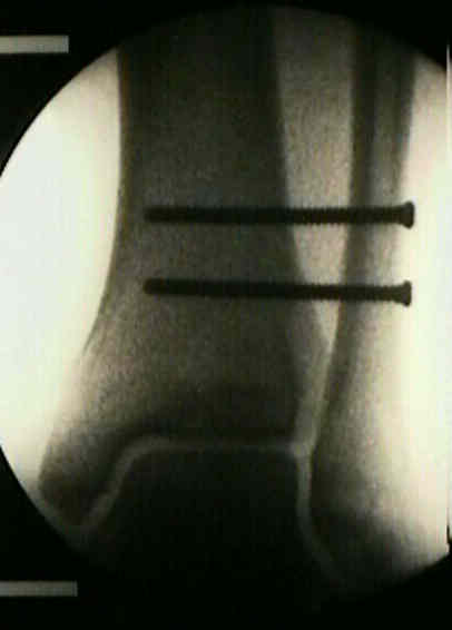
- Indications for Syndesmotic Fixation:
- combination of irreparable medial joint injury along w/ disruption of the syndesmosis > 4.5 cm proximal to the joint (Weber C Frx),
is an indication for surigical fixation;
- hence standard teaching is that w/ rigid ORIF of the medial malleolus frx, a syndesmotic screw would generally not be required);
- between 3 to 4.5 cm indication for fixation remains unclear;
- in contrast to the standard teaching, the report by Tornetta P (2000) demonstrated using an in vivo radiographic model that in bimalleolar fractures,
medial injury may be an osseous avulsion, leaving the deltoid intact on the displaced fragment, or it may be a combination of ligamentous
and osseous injury with disruption of the deep portion of the deltoid ligament;
- in the later case, ORIF of the medial malleolus may not restore function to the deltoid ligament;
- in this case, it is unclear whether syndesmotic fixation would be required;
- references:
- Competence of the Deltoid Ligament in Bimalleolar Ankle Fractures After Medial Malleolar Fixation
- in the report by Kennedy JG, et al, the authors examined the effect of syndesmotic screws in low Weber C fractures;
- low Weber C fractures are defined as being within 5 cm of the joint;
- 26 patients had ankle ORIF with syndesmotic fixation and 19 had ORIF w/o a syndesmotic screw;
- there was no significant difference between either group using subjective and objective criteria;
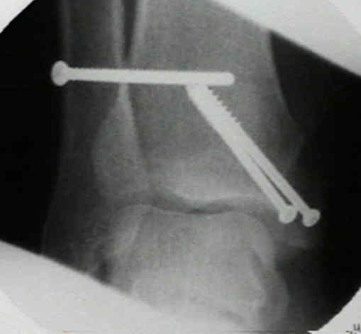
- high fibular frx / weber C Fractures:
- even w/ anatomic fixation of MM frx, syndesmotic screw fixation is indicated for fibular frxs occurring more than 15 cm above joint;
- syndesmotic fixation is recommeded w/ medial ligamentous injury, syndesmotic disruption, & talar shift w/o frx
 of fibula (diastasis);
- references:
- Evaluation of the syndesmotic screw in low Weber C ankle fractures.
- Ankle mortise stability in Weber C fractures. Indications for syndesmotic fixation.
- Contra-indications for syndesmotic fixation (w/ syndesmotic injury):
- w/ < 3 cm of sydesmotic disruption above plafond, there is little or no alteration was seen of ankle loading characteristics;
- anatomic reduction of fibula, esp if frx is w/in 4 cm of joint, tends to reduces talus in Mortise & restores syndesmotic stability;
- if medial malleolus is frxed & deltoid ligament is intact, ORIF of fibula & tibia should make syndesmosis fixation unnecessary;
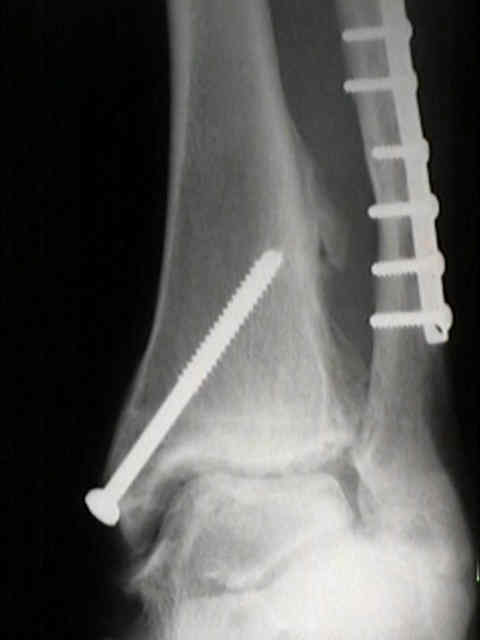
- case example:
- 40-year-old prisoner who sustained this Weber C frx which was fixed w/o a syndesmotic screw;
- 7 years later the patient had significant arthritis and significant pain;
- notice the heterotopic calcification along the interosseous membrane;
Competence of the Deltoid Ligament in Bimalleolar Ankle Fractures After Medial Malleolar Fixation
A radiographic evaluation of the tibiofibular syndesmosis.
Ligamentous injury of the lower tibiofibular syndesmosis: Radiographic evidence.
The fibular incisure of the tibia on CT Scan: a cadaver study.
Syndesmotic disruption in low fibular fractures associated with deltoid ligament injury.
Correlation of interosseous membrane tears to the level of the fibular fracture.
Radiographic Measurements Do Not Predict Syndesmotic Injury in Ankle Fractures: An MRI Study.
A biomechanical evaluation of clinical stress tests for syndesmotic ankle instability.