
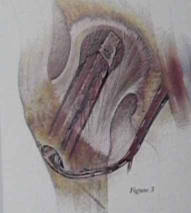
- Technique: When the lateral femur is exposed, a 3 mm guide pin is placed into the center of the necrotic area. Care must be taken, particularly on the lateral view, that there is enough room in the neck on both sides of the guide pin to pass a 21 mm reamer if needed. The leg can be flipped easily between the AP and lateral views at this time because the fibula graft should already be removed. The normal starting point for the guide pin is 2 cm distal to the lateral vastus ridge on the lateral femoral cortex. The guide pin is sometimes placed quite vertical in order to give support to the subchondral area of the defect. Cannulated reamers are progressively used over the guide pin starting with the 10 mm size. The average female and male require a final reaming diameter of 16 mm and 19 mm respectively. The size depends on the largest diameter of the fibular graft. The reaming extends to 3-5 mm from the articular surface. It is safer to do this reaming under live fluoroscopy. The bone from the reamers is saved for bone graft. The obviously necrotic bone from the femoral head is discarded. Once the optimal reaming size has been reached any further necrotic bone is removed under fluoroscope control with a ball reamer. The ball reamer is rarely used if the osteonecrosis is grade 3 or less. It is used to remove any cyst which can be seen on flouroscopy. Renograffin solution is instilled in the cavity to ensure the completeness of the removal.
