

- PreOp Planning:
- TKR Menu
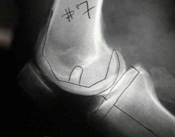
- Lateral View:
- note if AP size of lateral femoral condyle > medial condyle;
- w/ large discrepancy of small degree of Internal Rotation may be necessary to prevent undercutting anterior cortex;
- careful preoperative planning, using templates & lateral x-ray films, is critical to selecting the size of the femoral component;
- priority is given to reproduction of the A/P dimension, as this will restore normal kinematics and quadriceps muscle function;
- undersizing will result in looseness in flexion and possible anterior cortical notching of the femur;
- oversizing will create tightness in flexion and increased tension in quadriceps mechanism;
- Lateral Flexion View:
- w/ excessive gluide in flexion, PCL is probably tight, & therefore consider PCL sacrificing;
- Leg Alignment Radiographs:
- ensure that x-ray is not rotated:
- errors in rotation of radiographs can be diagnosed on x-ray by noting the normal relationship between the tibia
and the fibula at knee & at angle, as well as width of clear space between two bones;
- supine views:
- unstable joint external pressure upon the fully extended leg w/ pt supine and relaxed can easily change tibio-femoral angle;
- standing views:
- anatomic axis:
- mechanical axis:
- when standing normal leg in full extension is viewed from front, straight line connecting center of femoral head to center
of talus passes through center of the knee;
- Planning Femoral & Tibial Cuts:
- long term success of arthroplasty depends on restoration of the normal alignment of the lower limb;
- bringing transverse axis of the knee parallel to the ground in anatomic two legged stance;
- restoring normal wt. destribution across joint;
- ultimate result should place the transverse axis of prosthetic knee parallel to the ground as soon a possible;
- abnormalities in the coronal plane must be corrected accurately so that line of leg alignment passes, if possible, thru medial tibial
spine, but at least through middle third of knee joint;
- mechanical axis, & anatomic axis of the femur & tibia intersect at approx 7 deg of valgus;
- in the frontal plane, a line joining the tops of both tibial plateaus is oriented at approx 88 deg to anatomic-mechanical axis;
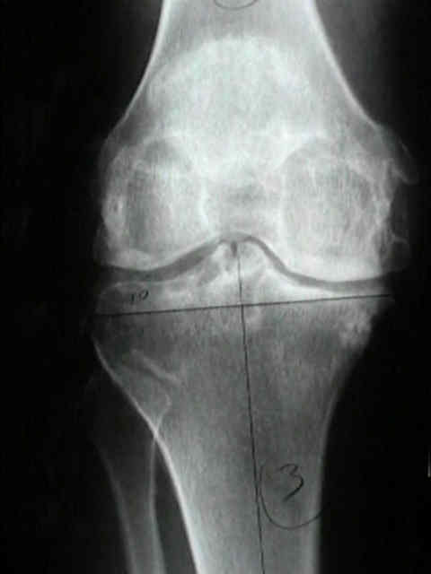
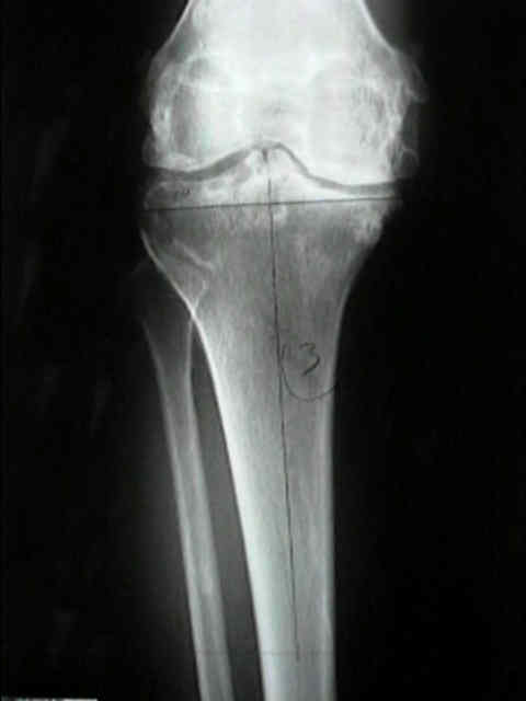
- Tibio-Fibular Angle:
- small variations in the tibio-femoral angle produce large changes in the position of the line of leg alignment;
- femoral compenent:
- should be placed in 9 +/- 2 deg of valgus from vertical axis;
- tibial component in 2-3 deg of varus;
- most recommend placing tibial component at 90 deg to anatomic mechanical axis;
- this position most closely approximates the average normal joint line during all phases of the gait cycle
Postoperative alignment of total knee replacement. Its effect on survival.
Alignment and long-term clinical results of a semiconstrained knee prosthesis.
Effect of knee component alignment on tibial load distribution with clinical correlation