


- Discussion:
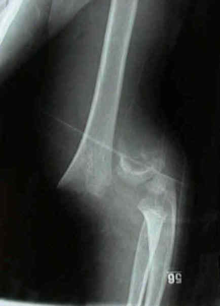
- posterior cortex is disrupted w/ no cortical contact;
- distal fragment is displaced posteriorly and proximally (by pull of triceps);
- w/ medial displacement, the medial periosteal hinge is intact;
- w/ lateral displacement, the lateral periosteal hinge is intact;
- Physical Exam:
- the proximal fragment tip may penentrate into the brachialis muscle;
- if the brachialis is buttonholed by the distal humeral spike, then the muscle can be milked off the spike by grasping the proximal arm and squeezing sequentially from proximal to distal;
- avoid excessive medial squeezing (to avoid N/V injury);
- references:
- Closed Reduction and Percutaneous Pinning of Displaced Supracondylar Humerus Fractures in Children: Description of a New Closed Reduction Technique for Fractures with Brachialis Muscle Entrapment.
- Radiographs:
- on AP view displacement may be posterolateral or posteromedial which has implications for both the reduction and surgical managment;
- adequacy of rotational alignment of distal fragment, is difficult to determine;
- rotation of distal fragment is best determined by CT;
- rotation of distal frag of > 10 deg results in a unacceptable varus deformity;
- Treatment:
- reduction;
- percutaneous pin fixation:
- displaced supracondylar frxs are reduced by closed methods & stabilized by percutaneous pin;
- this permits clinical evaluation of carrying angle once frx is stabilized;
- open reduction is indicated for difficult closed reduction (especially when the brachialis has button-holed through the brachialis)
Treatment of the displaced supracondylar fracture of the humerus (type III) with closed reduction and percutaneous cross-pin fixation.
Management of displaced extension-type supracondylar fractures of the humerus in children
Displaced fractures of the medial humeral condyle in children.
Transarticular fixation for severely displaced supracondylar fractures in children.
Year Book: Transarticular Fixation for Severely Displaced Supracondylar Fractures in Children.