

- See:
- TKR Menu:
- Collateral ligaments:
- Medial Collateral Ligament
- Lateral Collateral Ligament
- Discussion:
- more common in females;
- in valgus knee, ligament balancing is more difficult to fix;
- the anatomic deformity is usually larger in valgus than in varus, and it is on the femoral side;
- femur internally rotated, and tibia externally rotated;
- lateral orientation of tibial tubercle
- vastus lateralis enhanced as a dislocating moment of force on patella;
- lateral and posterior contractures, medial stretching
- typically the lateral femoral condyle is deficient;
- Problems:
- rotational mal-alignment of the AP cutting jig: (see: AP axis method);
- results from asymmetric wear of the posterior aspect of the lateral femoral condyle;
- posterior femoral condyles as a reference:
- w/ valgus deformity, there is defficiency of the of the posterior femoral condyle, which can lead to accidental internal rotation if the typical 3 deg
posterior condylar reference is used;
- in most knees the most reliable guide are the posterior condyles;
- w/ normal femoral anatomy, a slight relative external rotation (3 deg) of a line along posterior aspect of the femoral condyles will orient the AP
cuts perpendicular to the resected tibial surface;
- this technique, however, requires special care in any knee that has a large AP asymmetry in the femoral condyles;
- in such cases rotational alignment only w/ respect to posterior condyles may result in significant undercutting of anterior cortex laterally, severely notching cortex;
- when the posterior condyles are not available as a rotational reference (such as in AVN or severe valgus deformity), then the AP axis method should be used;
- patellar maltracking: (see: patellar subluxation)
- if significant preoperative valgus deformity has been corrected, it is often necessary to perform a lateral retinacular release to allow proper patellar tracking and
prevent patellar subluxation or dislocation;
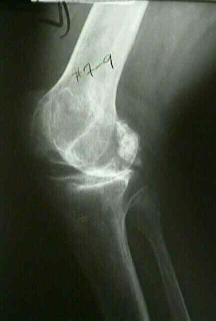
- if a patient has a preoperative valgus deformity of 7-9 deg, consider performing a distal femoral cut w/ 7 deg of valgus;
- in this case, it will rarely be necessary to perform a lateral release;
- peroneal nerve palsy:
- it is especially important that patients w/ significant valgus deformity not be given an epidural or a regional block when they undergo TKR;
- epidural anesthesia may contribute to peroneal nerve palsy in this situation, or alternatively it may mask a postoperative nerve palsy;
- if the initial postoperative exam reveals weak peroneal nerve function, then consider flexing the knee (which relaxes the nerve);
- instability
- especially posterolateral instability in flexion (see: flexion gap);
- need to have revision constrained components available;
- elevation of joint line:
- excessive valgus deformity w/ elongation of MCL requires extensive LCL and soft tissue release;
- extension gap increases when elongated medial structures and released lateral structures are balanced, thus requiring thicker tibia component and elevating joint line;
- average joint line elevation is approx 5.5 mm, which usually will cause no clinical performance of the patello-femoral joint;
- w/ the posterior stabilized prosthesis the joint line may be elevated up to 10 mm w/o problems;
- restoration of joint line is more critical in PCL retaining knees, in which PCL needs to be properly balanced in order to achieve good ROM;
- adjacent joint deformities:
- always note possible deformities in hip and foot prior to proceeding with knee arthroplasty;
- valgus foot puts a valgus strain upon the knee;
- deformity in the foot may be the cause of the deformity of knee in R.A.
- correction of ankle deformity is advised before reconstructive surgery of the knee is performed;
- Surgical Technique:
- surgical approach and general technique:
- proper alignment of femoral condyle
- rotational positioning of femoral component is important, for medial and lateral stability in both flexion and extension;
- lateral femoral condyle may be worn and atrophic on its distal and posterior surfaces;
- posteriorly the knee may be especiall worn, & aligning knee in standard fashion will put femoral component in internal rotation;
- answer is to line the cutting guides up with the AP axis and intercondylar notch or with the epicondyles, never with the posterior condyles;
- posteriorly, cut more medially than laterally, thickness of posterior cut should be much less on the lateral side than one medial side;
- soft tissue imbalance and releases for valgus deformity:
- all bony cuts are made prior to sequential soft tissue release;
- posterior stabilized trial prosthesis (with appropriate box cut and removal of PCL) helps determine degree of release;
- trial components and preparation for revision components:
- with proper ligament balancing, there still may be unacceptable laxity or instability;
- in this case, revision constrained components may be required
Correction of ligament and bone defects in total arthroplasty of the severely valgus knee.
The effect of extraarticular varus and valgus deformity on total knee arthroplasty.
The anteroposterior axis for femoral rotational alignment in valgus total knee arthroplasty.
The Difficult Knee: Severe Varus and Valgus.
Total Knee Arthroplasty for Severe Valgus Deformity. Five to 14-year follow-up.
Anatomy, Function, and Surgical Access of the Iliotibial Band in Total Knee Arthroplasty
Original Text by Clifford R. Wheeless, III, MD.
Last updated by Clifford R. Wheeless, III, MD on Sunday, February 3, 2013 8:26 pm