

- See:
- Revision TKR:
- TKR Menu
- Discussion:
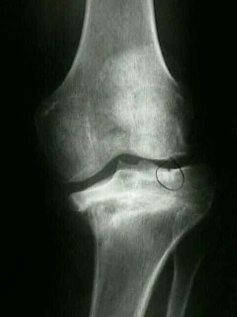
- asymmetrical bone loss often occurs in the proximal tibia;
- CT scan have shown that once one get more than 1 cm below joint line quality and quantity of the supporting cancellous bone
diminishes precipitously;
- equally important, attachments of iliotibial band, pes anserinus, patellar ligament, & PCL can be comprimised;
- problem of asymmetrical bone loss can be properly addressed in number of ways, including bone grafting, custom implants, or metal
wedges, or filling gap w/ cement (not first choice);
- Evaluation of Bone Defects:
- defects may be contained or non-contained
- uncontained defects:
- may be described as circumferential or non-circumferential, and whether the defect is greater than or less than 3 cm;
- bone loss of all, or almost all, of cancellous bone of metaphysis can also be present, leaving a funnel shaped cortical tube;
- defect of varus deformity is usually located on the periphery on posteromedial aspect of the tibia;
- valgus deformity more often produces contained posterolateral tibial defect in conjunction with a lateral femoral condylar deficiency;
- management:
- filling of defects w/ cement alone;
- cement w/ screw support;
- modular metal wedges, and custom components
- autogenous or allogenic bone graft;
- uncontained defects greater than 3 cm may be an indication for structural allografts;
- contained defects:
- goal is to obtain firm seating of the tibial tray on a rim of viable bone along w/ rigid press fixation of the medullary stem;
- Bone Defects in Primary Knees:
- Cementing of Bone Defects:
- methylmethacrylate cement is readily available, and its use is less technically demanding than other techniques;
- cement augmentation will work for small circumscribed areas w/ in intact peripheral rim, but will not support peripheral rim defects;
- cement filling should be limited to defects less than 1 cm;
- w/ defects > 1 cm use screws to act as reinforcement for the cement;
- consider limitined cement filling to defects < 1 cm;
- Bone Grafting of Defects:
- locally obtained autogenic bone graft usually is available in sufficient quantity during primary knee arthroplasty;
- fixation of these grafts requires the use of screws or pins, or the graft can be fashioned into a wedge to fit the tibia defect;
- intrusion of cement into the host-graft interface must be avoided because it impairs bone union.
- grafts that fill contained defects provide more reliable support for tibial component than do grafts that fill uncontained defects;
- goal is to obtain firm seating of the tibial tray on a rim of viable bone along w/ rigid press fixation of the medullary stem;
- structural bone grafts and morselized bone graft are more commonly used in revision arthroplasty, and are fixed with the same techniques described
for autologous grafts;
- long-term outcomes for allogenic graft are not yet available;
- complications:
- non union at the allograft-host bone junction;
- loosening w/ formation of radiolucent lines or migration of prosthesis;
- infection: may occur in about 12% of patients;
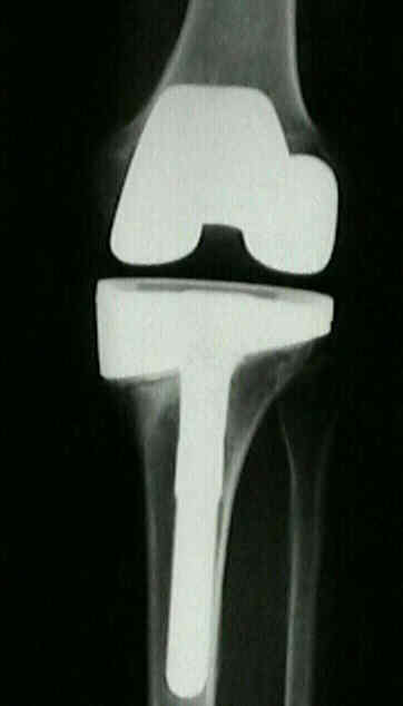
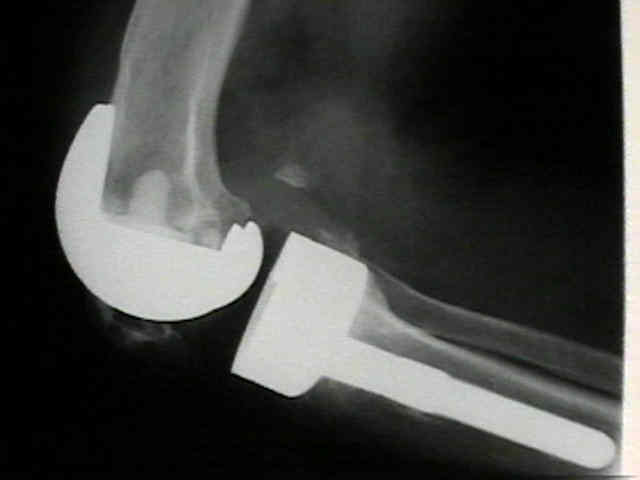
- Stems and Wedges:
- modular metal wedges have more stability than bone graft or cement;
- stem transfers up to 30% of force from bone subtending component to a point more proximal in the femur or more distal in the tibia;
- Case Example:
The incorporation of tibial allografts in total knee arthroplasty.
Inlay autogeneic bone grafting of tibial defects in primary total knee arthroplasty.
Correction of ligament and bone defects in total arthroplasty of the severely valgus knee.
Bone loss in the distal anterior femur after total knee arthroplasty.
The use of methylmethacrylate in primary total knee replacements with large tibial defects.
Autologous bone grafting for medial tibial defects in total knee arthroplasty.
Tibial wedge augmentation for bone deficiency in total knee arthroplasty. A followup study.
Radiologic and histologic analysis of morselized allograft in revision total knee replacement.
Original Text by Clifford R. Wheeless, III, MD.
Last updated by Clifford R. Wheeless, III, MD on Saturday, February 16, 2013 9:32 am