

- See: Infections of the Hand
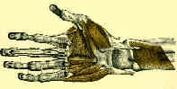
- Anatomy:
- extends mediolaterally from long metacarpal bone to the thenar eminence;
- extends proximodistally from transverse carpal ligament to just proximal to superfical palmar arch;
- borders of the thenar space include:
- adductor pollicis dorsally;
- index finger flexor tendons volarly;
- midpalmar septum ulnarly;
- thenar bursae minimizes friction between adductor pollicis & index finger tendons & thenar eminence;
- dorsal extension of the thenar bursa makes smoother the movements between adductor pollicis and the first dorsal interosseous, index metacarpal bone, and the first palmer interosseous;
- Tenosynovitis:
- suppurative tenosynovitis of index finger tendon sheath can rupture through its proximal end into the thenar bursae to cause thenar space abscess;
- thenar/mid palmar space infections;
- severe pain;
- swelling
- fullness of web space;
- abscess is deep to flexor tendons, and dorsal extension of abscess may be deep to the adductor pollicis;
- Treatment:
- consider using a combined palmar and dorsal approach to thenar space;
- palmar incision:
- make a transverse incision just proximal to MP flexion crease;
- avoid cutaneous nerves;
- w/ misquito clamp spread between metacarpals;
- dorsal incision:
- longitudinal incision is made on the dorsal aspect of the web between the thumb and index finger;
- spread to decompress the space behind the adductor pollicis and along the proximal radial aspect of 1st interosseous;
- closed suction irrigation:
- this technique has been used in that past, but is used less frequently at present because it is uncomfortable for the patient and time intensive for the nursing staff;
- technique:
- insert and secture 14 gauge catheter into palmar wound;
- loosely close palmar wound, and insert wick into dorsal wound;
- design bulky dressing so that it can be partially changed every shift;
- run D5LR at 50 cc/hour;
- second look in 48 hours