

- See: Role of the Ankle in Gait
- Discussion:
- is a hinge joint w/ malleoli projecting downward from tibia & fibula forming medial and lateral walls of the mortise, encompassing talus;
- in addition, malleoli serve as pulleys for tendons reaching plantar surface of foot from posterior and lateral compartments of leg.
- as is usual in hinge joints, capsular ligament is weak in front of & behind the joint and is reinforced by collateral ligaments at sides;
- on medial side, heavy deltoid ligament is attached above to medial malleolus and fans out to attach below chiefly to talus, but also to calcaneus;
- on lateral side there are three smaller ligaments: the anterior and posterior talofibular ligaments and calcaneofibular ligament, which lies between the talofibular ligaments;
- body and tibial articular surface of talus is wider anteriorly than posteriorly & form cone that is larger laterally than medially;
- due to this configuration, as ankle dorsiflexes intermalleolar distance increases slightly, talus externally rotates slightly, & fibula must laterally rotate slightly;
- slight rotary motion of the ankle in transvere plane causes the foot to adduct and abduct;
- these motions are coupled so that dorsiflexion and abduction occur at same time (plantar flexion & adduction occur at same time);
- since lateral malleolus is longer than medial malleoli & lies more posteriorly, ankle axis is accordingly angled both inferiorly & posteriorly;
- ROM in saggital plane is 20 deg of DF dorsiflexion to 50 deg PF;
- Ankle Joint and Subtalar Joint Work Together:
- ankle combines dorsiflexion w/ abduction & plantar flexion w/ adduction, subtalar joint combines dorsiflexion, abduction, & eversion in one direction and plantar flexion, adduction, and inversion in the other direction;
- these combined subtalar motions have traditionally been referred to as pronation & supination, respectively;
- when the subtalar joint is fused, rotation is increased in the ankle and may cause arthritic change;
- when the ankle is fused, greater stresses are placed on the sub-talar and midtarsal joints;
- Ankle Stability:
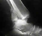
- see ankle sprains;
- stability of the ankle joint is based on the shaped of the articulation;
- best support is in dorsiflexion;
- 4 bony elements:
- medial malleolus and medial colateral ligaments;
- anterior syndesmotic ligaments and their bony attachment sites on tibia and fibula;
- posterior syndesmotic ligament & posterior malleolus;
- lateral malleolus and lateral collateral ligaments;
- fibula provides stability & preventing displacement of talus;
- shortened or malrotated fibula will allow talus to shift or tilt (see talar tilt) even if the medial ligaments are intact;
- even small changes significantly influence joint contact area;
- 1mm lateral shift of talus decreased contact area by 42%;
- 3 mm lateral shift decreases contact area decreased by > 60%;
- windlass action has been described in ankle, where full dorsflexion is limited by plantar aponeurosis & further tension on aponeurosis (e.g., with toe in dorsiflexion) will cause arch to rise;
- medial malleolus:
- medial malleolus is an extension of the distal tibia;
- inner surface is covered with articular cartilage and articulates with the medial facet of the talus;
- distal, inner surface of the malleolus is divided by a longitudinal groove into a large, anterior colliculus and a smaller, posterior colliculus, each an attachment site for portion of deltoid ligament;
- there is also a groove on the posterior surface where the posterior tibial tendon passes behind malleolus & tendon sheath is attached;
- Fibula:
- fibula provides the lateral support of the ankle;
- just above the ankle joint, the fibula sits in a groove formed by broad anterior tubercle & smaller posterior tubercle of the tibia;
- there is no articular surface between the distal tibia and fibula, even though there is a small amount of motion between these two bones;
- medial border of the fibula is covered by articular cartilage from level of the tibial plafond to a point approximately half way down its remaining length;
- distal end is tapered and has posteior goroove for the peroneal tendon;
- in lower limb, up to 1/6 of wt is carried by fibula & rest by tibia;
- fibula is pulled distally in stance phase by action of long toe flexors, interosseous membrane is tightened, mortise deepened, and the fibula pulled slightly medially, resulting in increase rotational stability of ankle;
- Interosseous Ligament:
- interosseous membrane runs between the tibia and fibula to level of the proximal tibiofibular joint;
- interosseous ligament is extension of interosseus membrane & is key transverse stabilizer of tibiofibular articulation;
- it stabilizes fibula, provides additional attachment sites for muscles, and may have some load bearing function
The contribution of the anterior talofibular ligament to ankle laxity.