

- Discussion:
- has become standard technique for stabilizing types II & type III frx;
- either two lateral pins, or one lateral and one medial pin may be used and both should penetrate the cortex;
- medial and lateral pin insertion provides better stabilization;
- 2 lateral pins may not permit full elbow extension, thus preventing full assessment of carrying angle;
- Radiographs:
- Baumann's Angle
- radiographs of uninjuried side
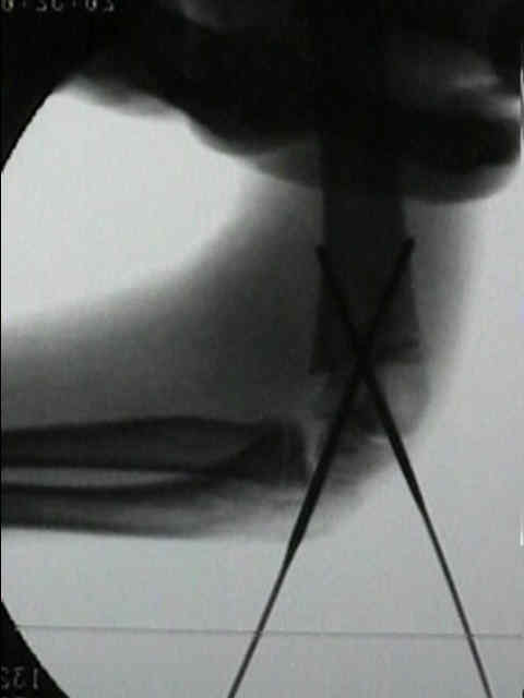
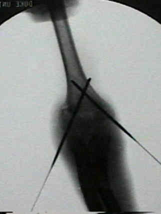
- Pinning Technique:
- reduction technique:
- in preparing for crossed pinning, keep elbow hyperflexed to maintain reduction;
- consider applying sterile "coband" to keep elbow flexed, which then allows arm to be externally rotated to achieve a lateral
view w/o moving flouro;
- pins should cross proximal to the frx at an angle of about 30 deg to the humeral shaft;
- positioning:
- w/ posteromedial displacement, place arm in maximum external rotation on flourscopy platform, and insert the medial pin first;
- w/ posterolateral displacement, place arm in maximum internal rotaiton on the flourscopy platform, and insert the lateral pin first;
- pin size:
- pins need to be smooth w/ trochar point;
- w/ children younger than 5-6 years, use 0.062 smooth K wire;
- in older children use 5/64 K wires;
- lateral pin:
- avoid directing pins too far anterior or posterior;
- Safe Zone for Superolateral Entry Pin Into the Distal Humerus in Children: An MRI Analysis
- insertion point is in the center of lateral condyle (capitellum);
- because the center of the capitellum is in line w/ anterior aspect of humeral shaft, the pin must be directed slightly posteriorly;
- wire is inserted thru the capitellum, and then the distal humeral physis;
- generally, the pin is aimed 35 deg upward and 10 deg posterior;
- pin should avoid the olecranon fossa and should come to rest along the far cortex;
- insert lateral pin first to obtain stability while reduction is evaluated (avoids need to repeatedly insert medial pins if reduction is
not adequate);
- consider placing a temporary 2nd pin thru the lateral condyle to achieve even more stability;
- Skaggs DL, et al:
- configuration of the pins did not affect the maintenance of reduction of either type-2 fractures or type-3 fractures;
- ulnar nerve injury was not seen in the 125 patients in whom only lateral pins were used;
- medial pin was associated w/ ulnar n injury in 4% patients in whom the pin was applied w/o hyperflexion of the elbow
and in 15% in whom the medial pin was applied w/ elbow hyperflexed;
- 2 years after the pinning, one of the 17 children with ulnar nerve injury had persistent motor weakness and a sensory deficit;
- crossed pin configuration:
- Cross pinning for supracondylar humerus fractures in children carries risk of iatrogenic ulnar nerve injuries
- Crossed Wires Versus 2 Lateral Wires in Management of Supracondylar Fracture of the Humerus in Children in the Hands of Junior Trainees.
- references:
- Loss of Pin Fixation in Displaced Supracondylar Humeral Fractures in Children: Causes and Prevention.
- Prevention of ulnar nerve injury during fixation of supracondylar frx by 'flexion-extension cross-pinning' technique.
- Biomechanical Analysis of Supracondylar Humerus Fracture Pinning for Fractures With Coronal Lateral Obliquity
- Biomechanical analysis of pin placement for pediatric supracondylar humerus fractures: does starting point, pin size, and number matter?
- Biomechanical testing of pin configurations in supracondylar humeral fractures: the effect of medial column comminution.
- Lateral-Entry Pin Fixation in the Management of Supracondylar Fractures in Children.
- Three lateral divergent or parallel pin fixations for the treatment of displaced supracondylar humerus fractures in children.
- A prospective randomised, controlled clinical trial comparing medial and lateral entry pinning with lateral entry pinning for percutaneous fixation of displaced extension type supracondylar fractures of the humerus in children.
- Intraoperative Stability Testing of Lateral-Entry Pin Fixation of Pediatric Supracondylar Humeral Fractures
- posterior intrafocal pin:
- Treatment of Gartland Type III Pediatric Supracondylar Humerus Fractures with the Kapandji Technique in the Prone Position.
- Biomechanical Analysis of Posterior Intrafocal Pin Fixation for the Pediatric Supracondylar Humeral Fracture
- The posterior intrafocal pin improves sagittal alignment in Gartland type III paediatric supracondylar humeral fractures.
- medial pin:
- passed obliquely through medial epicondyle, just proximal to olecranon fossa;
- need to protect ulnar nerve;
- note that w/ flexion, the ulnar nerve can sublux over the medial condyle placing it at risk w/ medial pin insertion;
- because of ulnar nerve subluxation, some surgeons always place the lateral pin first (w/ elbow hyperflexed) which confers
stability;
- once the lateral pin has been inserted, the surgeon can then bring the elbow out to 80-90 deg flexion (decreasing ulnar nerve
subluxation) prior to placement of the medial pin;
- surgeon's thumb can milk the ulnar nerve back into its posterior position and hold it there;
- if excessive soft tissue swelling is present, then consider making a small incision thru the skin over the medial epicondyle,
and then spreading w/ hemostat;
- use soft tissue protector from the cannulated screw set inorder to further protect the ulnar nerve;
- references:
- Iatrogenic ulnar nerve injury after treatment of supracondylar fractures: number needed to harm, a systematic review.
- Treatment of displaced supracondylar frx patterns requiring medial fixation: a reliable and safer cross-pinning technique.
- Is Medial Pin Use Safe for Treating Pediatric Supracondylar Humerus Fractures?
- Paediatric supracondylar frx: technique for safe medial pin passage with zero incidence of iatrogenic ulnar nerve injury.
- because medial epicondyle is slightly posterior to the shaft, direct the medial pin slightly anterior;
- also ensure that the medial pin enters straight into the epicondyle rather than distal to the epicondyle;
- the medial wire will often appear more transverse than the lateral pin;
- in the report by Skaggs DL, et al:
- use of a medial pin was associated w/ ulnar n injury in 4% patients in whom the pin was applied w/o hyperflexion of the
elbow and in 15% in whom the medial pin was applied w/ elbow hyperflexed;
- 2 years after the pinning, 1 of 17 children with ulnar nerve injury had persistent motor weakness and a sensory deficit;
- authors note that if a medial pin is used, the elbow should not be hyperflexed during its insertion;
- Loss of Pin Fixation in Displaced Supracondylar Humeral Fractures in Children: Causes and Prevention.
- after pin placement assess carrying angle to r/o cubitus varus
- Baumann's angle, angle between long axis of humeral shaft & growth plate of capitellum, will suggest final carrying angle after
reduction;
- finally, re-check the radial pulse and the quality of the pulse;
- casting:
- Immobilization After Pinning of Supracondylar Distal Humerus Fractures in Children: Use of the A-frame Cast
- Factors Affecting Forearm Compartment Pressures in Children with Supracondylar Fractures of the Humerus
- Post Op:
- pins can usually be removed a 3 weeks post op;
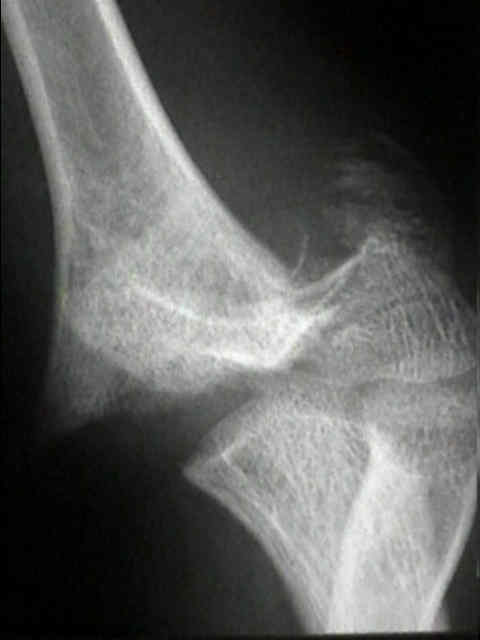
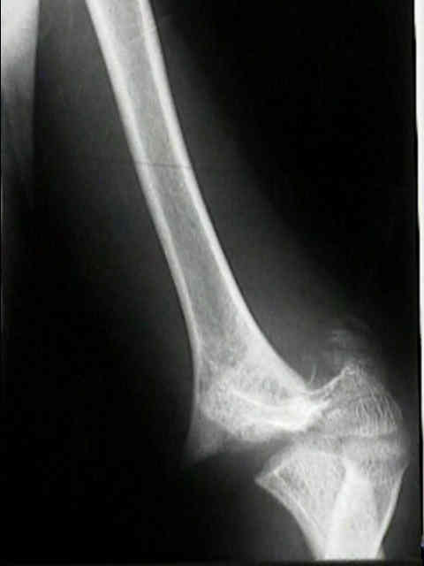
- Case Example:
- 7-year-old female who presented with a displaced supracondylar fracture
- an attempt at closed reduction was carried out, but the reduction was unacceptable;
- open reduction was carried out via limited medial and lateral incisions
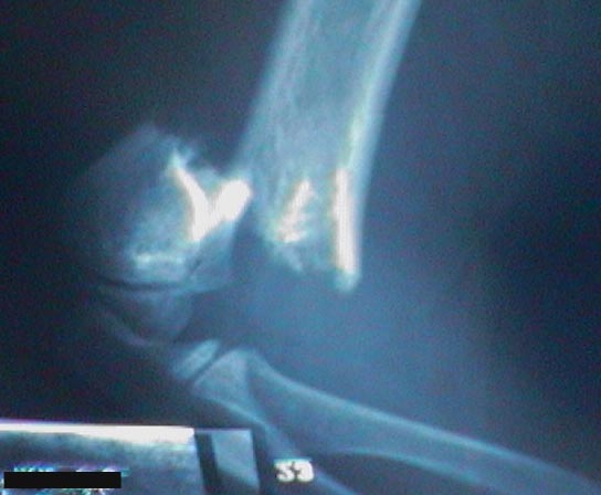
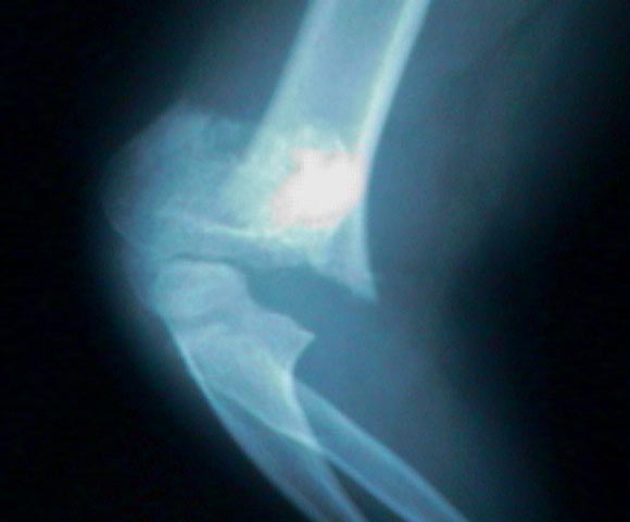
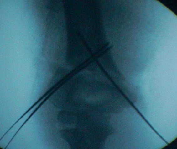
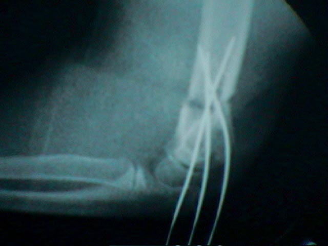
- Example:
- these pictures show residual displacement following closed reduction and pin fixation
Supracondylar fractures of the humerus in children. A modified technique for closed pinning.
Percutaneous fixation of supracondylar fractures of the humerus in children.
Difficult supracondylar elbow fractures in children: analysis of percutaneous pinning technique.
Supracondylar fractures of the humerus: a prospective study of percutaneous pinning.
Reduction and pinning of pediatric supracondylar humerus fractures in the prone position.