

 - See: Posterior Approach for Pelvic Frx
- Indications for Use:
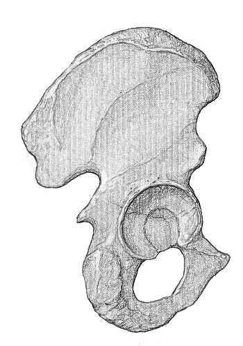
- isolated posterior wall & posterior column frxs;
- allows access to posterior column and posterior wall only, but exposure is limited proximally by
superior gluteal vessels and greater trochanter;
- isolated transverse frx (as well as associated transverse frxs and posterior wall frx);
- T shaped fractures (may also use extended iliofemoral approach);
- PreOp Planning:
- foley catheter;
- distal femoral traction pin;
- consider use of EMG w/ needles placed in TA and P longus (asseses peroneal div of
sciatic nerve) and needles placed in A hallucis and FHL muscles (which are innervated
by the posterior tibial nerve);
- spontaneous EMG allows real time monitoring of potenital sciatic palsy;
 - Positioning:
 - options include prone positioning, lateral positioning, or lateral positioning on the fracture table;
- some surgeons prefer the lateral position for posterior wall fractures and prone position for
posterior column fractures;
- avoid injury to the sciatic nerve;
- to protect the nerve the knee must be in flexion at all times
- Exposure:
- mark out PSIS, sciatic notch, and greater trochanter;
- incision:
- incision begins lateral to PSIS, crosses most anterior portion of notch, and subsequently crosses
posterior 1/3 of greater tuberosity;
- incise the skin, subQ tissues, and tensor fascia lata & blunting split the fibers of the maximus;
- excessive proximal spliting of the maximus may injure the inferior gluteal nerve;
- release a portion of gluteal sling for additional exposure (noting that the major insertion of the maximus is to IT band);
- trochanteric bursa is incised from distal to proximal, which helps to identify the posterior edge of gluteus medius and places
surgeon at correct plane for identifying the sciatic nerve;
- sciatic nerve is identified: (see protection of sciatic nerve in THR)
- at this point, identify: piriformis, quadratus femoris, & sciatic nerve;
- sciatic nerve is identified on the superficial surface of the quadratus muscle;
- note any contussions or discolorations of the nerve;
- to protect the nerve the knee must be in flexion at all times;
- carefully mobilize the sciatic nerve from its bed of areolar tissue and pass a penrose drain around it for identification;
- a nerve stimulator may be used to ensure that both divisions of the nerve are contained in the penrose drain;
- the hip remains extended and the knee flexed through out the procedure inorder to protect the nerve;
- gluteus maximus split:
- maximus has dual blood supply which is therefore tolerant of dissection;
- innervation is derived from inferior gluteal nerve and hence no internervous plane;
- hence muscle fibers are split up until the first nerve branch to the upper part of the muscle is seen;
- partially release the release of the gluteus maximus insertion into the femur, which allows adequate posteromedial
retraction of the maximus without stretching of the inferior gluteal nerve
- superior elevation of the medius:
- identify the interval between the gluteus medius and the piriformis;
- elevate the gluteus medius (off the pelvis) and retract it superiorly;
- then drive a large Steiman pin into the ilium at a point well above the acetabulum, which will keep medius retracted
superiorly throughout the case;
- reflect short external rotators:
- develop plane between the external rotators and underlying hip capsule (w/ periosteal elevator inserted just above
piriformis and directed distally);
- piriformis and the conjoined tendon (gemelli and the obturator internus) are tagged for later repair;
- be careful not to dissect around or injury the quadratus so as to avoid injury to the MFCA;
- incise external rotators about 1.5 cm from their insertion points into the greater trochanter inorder to avoid MFCA;
- reflect short external rotators off of their insertion and reflect them posteriorly inorder to protect the nerve;
- note that in some cases the external rotators can be partially avulsed from their origin;

- greater sciatic and lesser sciatic notch:
- identify the greater and lesser sciatic notch;
- injury to superior gluteal artery & nerve must be avoided;
- they can be visualized exiting from greater sciatic notch;
- exposure of the ilium;
- exposure of ilium will be limited because superior gluteal artery & nerve enter medius and minimus
limiting upward mobilization;
- elevate the gluteus medius and minimus muscle origins from the external surface of the ilium
- once the greater sciatic notch is adequately exposed, insert a curved homan retractor;
- the reflected external rotators should protect the sciatic nerve from the Homan;
- a second homan rectrator is placed into the lesser sciatic notch, just below the ischial spine;
- take care not to injure the pudenal artery;
- this will help retract the conjoined muscles and the sciatic nerve posteriorly;
- greater trochanteric osteotomy:
- patients undergoing osteotomy may be at greater risk for heterotopic ossification;
- sliding osteotomy may be procedure of choice;
- this type of osteotomy facilitates visualization of the superior aspect of the hip capsule;
- performed correctly, this type of osteotomy should not interfere w/ MFCA and blood supply to the hip;
- distal end of trochanter is left attached to the vastus lateralis and the proximal end attached to the
gluteus medius and minimus;
- references:
- Osteotomy of the Trochanter in Open Reduction and Internal Fixation of Acetabular Fractures.
- Direct complications of trochanteric osteotomy in open reduction and internal fixation of acetabular fractures.
- Trochanteric flip osteotomy for cranial extension and muscle protection in acetabular fracture fixation using a Kocher-Langenbeck approach.
- The role of trochanteric flip osteotomy in fixation of certain acetabular fractures.
- other measures to improve exposure:
- origin of the hamstrings can be elevated from the ischial tuberosity to expose the lower posterior column;
- hip capsule can be released from the intact portion of the acetabular rim (perimeter of the labrum);
- these measures may allow for visualization of the femoral head (after hip dislocation), femoral head debridemont, and
management of associated fractures (such as transverse fracture or posterior wall fractures);
- Deep Exposure:
- exposure of posterior column:
- entire posterior column of the acetabulum is exposed; using blunt dissection, elevate medius from outer side of ilium;
- consider femoral distractor:
- consider trochanteric osteotomy:
- if visualization of superior dome and anterior column is needed;
- for difficult transverse or T type frxs;
- exposure of the posterior wall: (see: posterior wall)
- determine whether there is any pre-existing posterior capsular stripping;
- posterior capsulotomy is performed by detaching it from its acetabular origin;
- detachment from the femoral origin may disrupt the blood supply to the femoral head, which could lead to AVN;
- quadrilateral surface:
- may be palpated through the greater or lesser sciatic notch;
- dislocation of hip:
- indicated for interposed intra-articular fragments;
- consider sliding trochanteric osteotomy for easier dislocation;
- consider intra operative drilling of the femoral head inorder to demonstrate perfusion;
- reference:
- Surgical dislocation of the hip for the fixation of acetabular fractures
- Complications:
- sciatic nerve palsy:
- most common cause of palsy is retraction of sciatic nerve;
- to avoid palsy, keep patient's knee flexed at least 60 deg & hip extended;
- if sciatic nerve palsy occurs, it is treated initially with an AFO,
- there may be improvement in the palsy for up to 3 years
Femoral artery thrombosis after open reduction of an acetabular fracture.
Long-term results in surgically treated acetabular fractures through the posterior approaches.