

- Pathogenesis:
- see: hematogenous osteomyelitis and pediatric bone circulation
- tortuous course of the nutrient vessels in bone causes bacteria to be trapped in the metaphysis;
- once lodged in the metaphysis, the infection spreads toward the epiphysis;
- in neonates the periosteum is loosely applied to the cortical surface, which can allow pus to track upwards between these surfaces and extend over the length of the bone or rupture into the soft tissues;
- Associated Conditions:
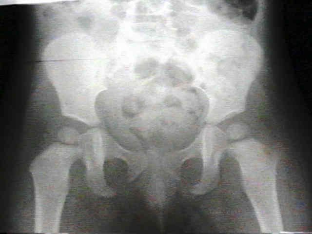
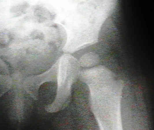
- septic arthritis:
- septic arthritis is both more common & more often assoc w/ metaphyseal osteomyelitis in neonates than in older children;
- in some series, 60-100% of neonates with septic arthritis have adjacent osteomyelitis;
- note that multiple sites of involvement are common in infants (consider ultrasound if there is additional large joint tenderness)
- epiphyseal plate prevents the infection from entering the joint space in older children but apparently does not act as a barrier in infants;
- these transphyseal blood vessels are thought to disappear by age 6 mo, but the exact age of this transition remains unclear;
- ref: The arterial supply of the developing proximal end of the human femur.
- joint infection secondary to osteomyelitis may occur in shoulder and hip as result of the synovial membrane inserting distally to epiphysis, allowing bacteria to spread directly from the metaphysis to joint space;
- only metaphysis of shoulder, hip, radial head, and ankle remain intracapsular during early childhood;
- the hip joint seems especially prone to sepsis from adjacent osteomyelitis (see septic hip);
- these synovial reflections over the metaphyseal bone decrease with age;
- Clinical Findings:
- deep soft tissue swelling may not be early x-ray feature because neonates lack subQ fat and have poorly defined fascial planes;
- at knee, joint is usually spared, and infections beginning in distal femoral or proximal tibial metaphysis usually spread along shaft of bone moving away from joint;
- tenderness over overlying muscles may indicate that the pus has ruptured through the periosteum and into the soft tissues;
- Radiographic Studies:
- many feel that bone scans are a poor choice in infants < 6 months, with negative studies in upto 60% of cases of OM
- Management:
- aspiration of site:
- in order to recover causitive organisms & to determine whether an abscess is present, which wound require surgical drainage;
- aspiration is essential if a bone abscess or unusual organism is suspected;
- treatment:
- neonates
- ages from 6 months to 2 years
- criteria for oral ATBs
The futility of bone scanning in neonatal osteomyelitis: concise communication.
- see: hematogenous osteomyelitis and pediatric bone circulation
- tortuous course of the nutrient vessels in bone causes bacteria to be trapped in the metaphysis;
- once lodged in the metaphysis, the infection spreads toward the epiphysis;
- in neonates the periosteum is loosely applied to the cortical surface, which can allow pus to track upwards between these surfaces and extend over the length of the bone or rupture into the soft tissues;
- Associated Conditions:
- septic arthritis:
- septic arthritis is both more common & more often assoc w/ metaphyseal osteomyelitis in neonates than in older children;
- in some series, 60-100% of neonates with septic arthritis have adjacent osteomyelitis;
- note that multiple sites of involvement are common in infants (consider ultrasound if there is additional large joint tenderness)
- epiphyseal plate prevents the infection from entering the joint space in older children but apparently does not act as a barrier in infants;
- these transphyseal blood vessels are thought to disappear by age 6 mo, but the exact age of this transition remains unclear;
- ref: The arterial supply of the developing proximal end of the human femur.
- joint infection secondary to osteomyelitis may occur in shoulder and hip as result of the synovial membrane inserting distally to epiphysis, allowing bacteria to spread directly from the metaphysis to joint space;
- only metaphysis of shoulder, hip, radial head, and ankle remain intracapsular during early childhood;
- the hip joint seems especially prone to sepsis from adjacent osteomyelitis (see septic hip);
- these synovial reflections over the metaphyseal bone decrease with age;
- Clinical Findings:
- deep soft tissue swelling may not be early x-ray feature because neonates lack subQ fat and have poorly defined fascial planes;
- at knee, joint is usually spared, and infections beginning in distal femoral or proximal tibial metaphysis usually spread along shaft of bone moving away from joint;
- tenderness over overlying muscles may indicate that the pus has ruptured through the periosteum and into the soft tissues;
- Radiographic Studies:
- many feel that bone scans are a poor choice in infants < 6 months, with negative studies in upto 60% of cases of OM
- Management:
- aspiration of site:
- in order to recover causitive organisms & to determine whether an abscess is present, which wound require surgical drainage;
- aspiration is essential if a bone abscess or unusual organism is suspected;
- treatment:
- neonates
- ages from 6 months to 2 years
- criteria for oral ATBs
The futility of bone scanning in neonatal osteomyelitis: concise communication.
Osteomyelitis in the neonate.
Regeneration of the proximal tibial epiphysis after infantile osteomyelitis: report of three cases with an eight- to 22-year follow-up.
Clinical and diagnostic features of osteomyelitis occurring in the first three months of life.