

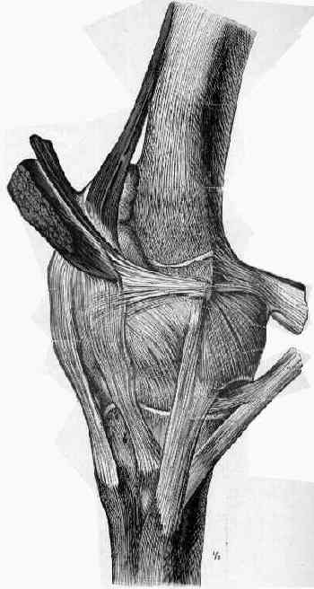
- medial patellofemoral ligament, a distinct condensation of capsular fibers in coronal plane which originates at
the medial epicondyle (adductor tubercle and adductor tendon) as well as the MCL;
- deep fascia and aponeurotic edge of the VMO fuses with the superior border of the medial
patellofemoral ligament;
- it runs transversely (deep to the distal VMO) and inserts on superomedial aspect of patella;
- helps resist lateral migration of patella (provides 50%-80% of the restraining force to lateral patella dislocation);
- MPFL is most effective between 0 and 30 deg flexion (trochlea is primary restraint with additional flexion) ;
- relation to medial knee compartments:
- there are three anatomic layers on the medial side of the knee;
- superficial layer: investing fascia over the sartorious muscle;
- middle or second layer: includes the parapatellar retinaculum, MPFL, and superficial MCL;
- third or deep layer: includes the deep medial collateral and joint capsule;
- references:
- The anatomy and reconstruction of the medial patellofemoral ligament.
- Evaluation of the medial soft-tissue restraints of the extensor mechanism of the knee.
- Anatomy and biomechanics of the medial patellofemoral ligament.
- Medial soft tissue restraints in lateral patellar instability and repair.
- Patellar Subluxation at Terminal Knee Extension: Isolated Deficiency of the Medial Patellomeniscal Ligament
- The Relationship of the Medial Patellofemoral Ligament Attachment to the Distal Femoral Physis
- Rupture:
- rupture of medial patellofemoral ligament may follow lateral dislocation of the patella;
- ligament usually ruptures from its femoral origin;
- pain and tenderness along the medial retinaculum;
- need to distinguish from MCL tear;
- ref: Traumatic Patellar Dislocation and Clinical Significance of Medial Patellofemoral Ligament Injury
- Surgical Repair: (for femoral avulsion);
- MRI is useful to confirm femoral avulsion (versus the occassional patellar avulsion);
- 4 cm longitudinal incision made anterior to medial epicondyle at the edge of the VMO;
- MPFL is identified below level of muscle fascia;
- ligament is re-attached to the femoral using bone anchors;
- Surgical Reconstruction:
- gracilis or semitendenosis tendon harvest: (see hamstring harvest for ACL reconstruction)
- tendon is harvested through one inch incision over the pes anserinus;
- gracilis tendon is exposed and released with a tendon stripper
- patellar attachment:
- ref: The Patellar Insertion of the Medial Patellofemoral Ligament in Children: A Cadaveric Study
- femoral attachment:
- radiographically proximal height of the femoral tunnel is at superior 1/3 of patella with the knee in 20-30 knee flexion;
- ideally located at the epicondyle or mid way between the epicondyle and the adductor tubercle:
- adductor tubercle is exposed and identified just proximal to the medial femoral condyle;
- MPFL insertion point is slightly distal to the tubercle
- references:
- Changes in the length of the medial patellofemoral ligament: an in vivo analysis using 3-dimensional computed tomography.
- The Relationship of Femoral Tunnel Positioning in Medial Patellofemoral Ligament Reconstruction on Clinical Outcome and Postoperative Complications.
- avoid proximal and anterior malposition
- biggest mistake is insertion of femoral tunnel too proximal and too anterior;
- proximally malpositioning the femoral attachment point of the reconstructed MPFL will increase the force and pressure
applied to cartilage on the medial facet of the patella;
- subcuntaneous passage vs subfascial passage:
- subcutaneous passage allows ligamentous reconstruction without disrupting the remants medial patellofemoral ligament;
- deep passage of the graft (just above the capsule) risks allowing the graft to "cheese cutter" down over the capsule and
impinge on the medial femoral condyle during flexion;
- references:
- The role of medial retinaculum plication versus medial patellofemoral ligament reconstruction in combined procedures for recurrent patellar instability in adults.
- Medial patellofemoral ligament reconstruction using semitendinosus tendons: polyester suture augmentation versus nonaugmentation.
- Comparison Between a Static and a Dynamic Technique for Medial Patellofemoral Ligament Reconstruction
- Medial retinaculum plasty versus medial patellofemoral ligament reconstruction for recurrent patellar instability in adults: a randomized controlled trial.
- Medial patellofemoral ligament reconstruction with semitendinosus autograft for chronic patellar instability: a follow-up study.
- A simple technique for reconstruction of the medial patellofemoral ligament using a quadriceps tendon graft.
- Reconstruction of the Medial Patellofemoral Ligament Using the Adductor Magnus Tendon: An Anatomic Study
- Medial patellofemoral ligament reconstruction as an isolated or combined procedure for recurrent patellar instability.
- Reconstruction of the Medial Patellofemoral Ligament Using a Quadriceps Tendon Graft: A Case Series
- Medial Patellofemoral Ligament Reconstruction With Half Width (Hemi Tendon) Semitendinosus Graft
- lateral retinacular release:
- indicated when there is residual lateral subluxation;
- tensioning of the graft:
- may be carried out in 30-40 deg of knee flexion;
- careful not to overtension;
- Complications:
- saphenous neuritis: (see saphenous nerve)
- Gracilis transfer associated with distal alignment for patella alta with recurrent dislocations: An original surgical technique
- Reconstruction of the Medial Patellofemoral Ligament Using the Adductor Magnus Tendon: An Anatomic Study
- references:
- Technical Errors During Medial Patellofemoral Ligament Reconstruction Could Overload Medial Patellofemoral Cartilage.
- Technical Failure of Medial Patellofemoral Ligament Reconstruction
- Primum non nocere: MPFL reconstruction complications
- Analysis of failure and clinical outcome after unsuccessful medial patellofemoral ligament reconstruction in young patients.
Reconstruction of the medial patellofemoral ligament for the treatment of habitual or recurrent dislocation of the patella in children.
Results of medial patellofemoral ligament reconstruction in the treatment of patellar dislocation.
Semitendinosus tenodesis for repair of recurrent dislocation of the patella in children.
Medial patellofemoral ligament reconstruction with semitendinosus autograft for chronic patellar instability: a follow-up study.
Reconstruction of the Medial Patellofemoral Ligament With Gracilis Tendon Autograft in Transverse Patellar Drill Holes
In vitro investigation of the effect of medial patellofemoral ligament reconstruction and medial tibial tuberosity transfer on lateral patellar stability.
Reconstruction of medial patellofemoral ligament for chronic patellar instability
Medial Patellofemoral Ligament Reconstruction With Half Width (Hemi Tendon) Semitendinosus Graft
MPFL reconstruction for patellar instability in younger patients: Save it for the recurrent dislocators
Medial patellofemoral ligament reconstruction for patellar instability in patients with hypermobility: A case control study