

- See: Medial Collateral Ligament:
- Superficial Layer (Layer I)
- most superficial layer includes the deep fascia that arises from anterior to posterior encases entire medial aspect of
the knee & coalesces w/ hamstring muscles and posteromedial capsule;
- this first layer is superficial to superficial MCL;
- includes investing fascia of sartorius, fascia overlying gastroc & popliteal fossa;
- anteriorly layer I blends w/ medial retinaculum as it inserts into proximal aspect of the tibial palteau;
- superficial fascia layer of medial retinaculum of knee joints posteromedially with layer of the pes arserinus tendons
(including semitendinosus) and continues posteriorly as a common sheath in popliteal fossa;
- Middle Layer (Layer II)
- includes superficial MCL which has very distal insertion on medial aspect of tibia at level of pes anserinus;
- considered the primary static stabilizer to valgus stress;
- there may be both parallel & oblique portions of this MCL, which inserts almost 8 cm below joint line & is post to pes anserinus;
- most posterior portion of layer II, from posterior oblique ligament, has direct fibers that run from medial epicondyle, blending w/layer III &
attaching to posterior tibial articular surface;
- these fibers are also augmented by contricutions from fascia overlying semimembranous;
- anteriorly, layer II splits vertically, w/ one portion merging w/ medial retinaculum & other w/ patellofemoral ligament;
- Deep Layer:
- deep MCL (deepest layer of medial structures)
- its posterior extension, posteromedial capsule, includes posterior oblique ligament, described by Hughston;
- true capsule of joint is most intimate connection w/ synovium;
- it is very thin anteriorly, but at level of joint, deep to superficial MCL, is thickening of layer III identified as deep MCL;
- anteriorly, there is bursa interposed between these 2 ligaments, whereas posteriorly they merge w/ ea other & meniscotibial lig;
- deep MCL is a major secondary restraint to anterior translation
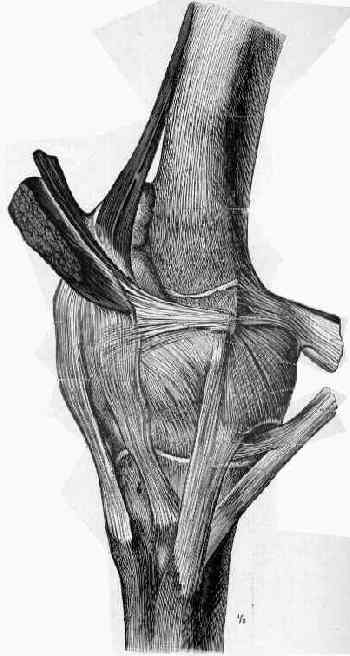
The supporting structures and layers on the medial side of the knee: an anatomical analysis.