- Discussion:
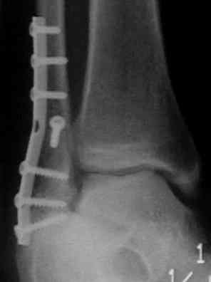
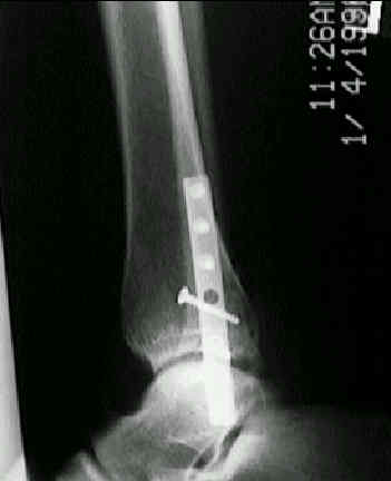
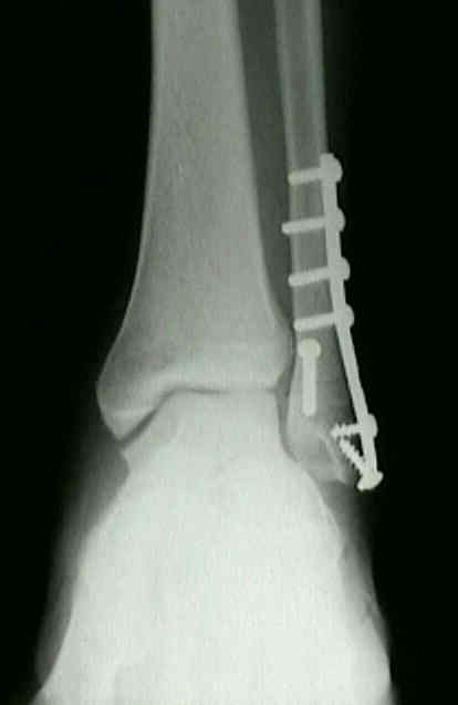
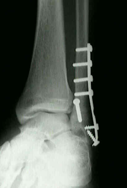
- when the frx is sufficiently oblique & is not comminuted, it can be treated w/ a lag screw to produce intrafragmentary compression, and neutralization plate placed laterally to prevent frx from slippling;
- disadvantages of lateral plating:
- prominent lateral screws may cause symptoms or wound necrosis;
- possibility of distal intra-articular screw insertion, and on the contrary, there is the possibility of inadequate fixation if distal screws are too short;
- may not allow adequate fixation in osteoporotic bone;
- may interfere w/ syndesmotic screw insertion (especially when two syndesmoic screws are to be used);
- preoperative considerations:
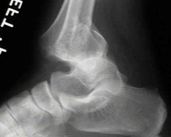
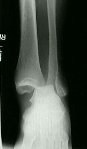
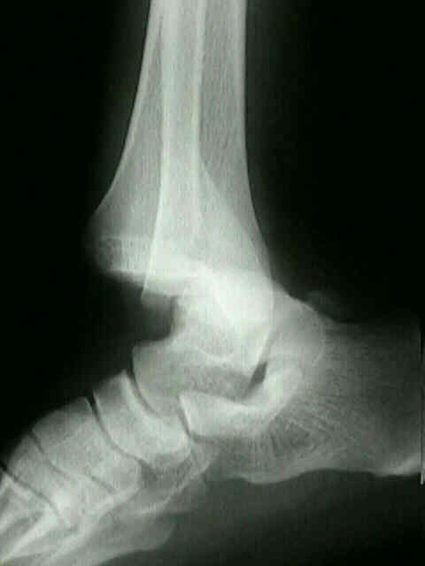
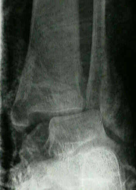
- fracture dislocations of the ankle
- open ankle fractures
- trimalleolar frx (posterior malleolar frx);
- syndesmotic injury:
- consider ahead of time how and where the syndesmotic screw(s) will transverse the plate;
- alternattive fixation techniques:
- fixation w/ two lag screws
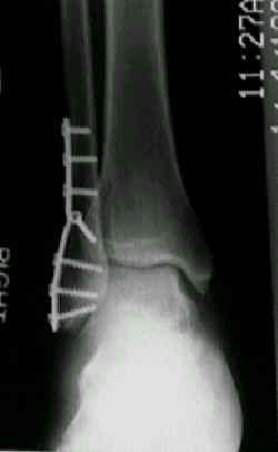
- posterior antiglide plate
- supplemental K wire fixation:
- w/ significant osteopenia, there may be a higher incidence of hardware failure;
- consider use of preliminary 0.62 K wires which are inserted from the tip across the frx to penetrate the medial cortex of the proximal fragment;
- one wire is placed anteriorly and one is placed posteriorly in order to allow insertion of plate screws between the K wires;
- the distal exposed K wires are bent and cut;
- ref: A new technique for complex fibula fracture fixation in the elderly: a clinical and biomechanical evaluation.
- Operative Technique:
- patient position:
- supine, tourniquet, hip bump to internally rotate the leg, and pelvic strap to allow tilting of table if needed;
- flouroscopy is required if there is a syndesmotic injury or if there is extensive comminution (so that reduction of fibula can be judged based on the congruency with the talus);
- medial malleolus fracture:
- should be fixed first because the anatomic reduction is easy, and helps guide the reduction of the fibulur frx;
- 4.0 mm cancellous bone screws, or 4.0-4.5 mm cannulated screws are used for fixation of medial malleolus & posterolateral tibial fragment;
- alternatively use K wires, 1.6 mm diameter, and 1.2 mm cerclage wires for tension band wiring of the medial malleolus;
- exploration of ankle joint:
- need to look for osteochondral fragments;
- after exposure of frx & anterior surface of fibula, joint is explored;
- this should be repeated during the approach to the lateral malleolus;
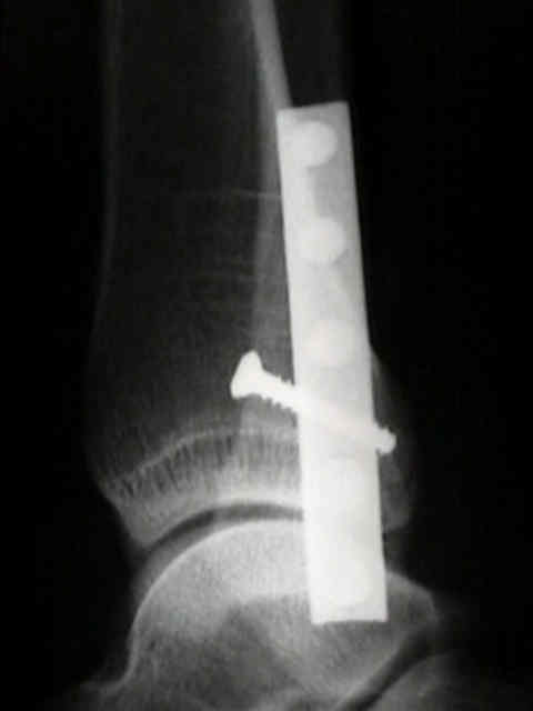
- lateral malleolus frx
- 3.5 mm cortex screws are used as lag screws in oblique fractures of the fibula;
- screws must engage posterior cortex for secure fixation but should not protrude far enough to encroach on peroneal tendon sheaths;
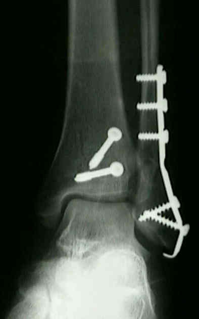
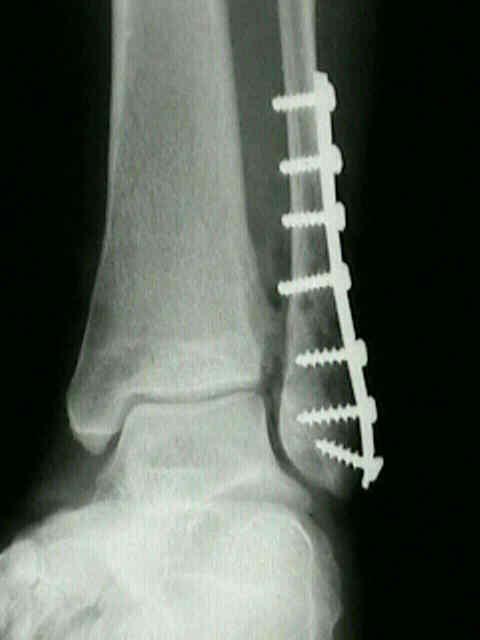
- one third tubular plate is applied to the lateral malleolus, using 3.5 mm cortical screws proximally and 4.0 mm cancellous screws distally;
- surgical approach of lateral malleolus
- fracture reduction
- comminution:
- w/ extensive comminution is can be difficult to achieve reduction and it is difficult to know when the frx is out to length;
- failure to restore normal length & rotation of fibula often leads to a poor result;
- fibula is brought out to length and the reduction is judged based on the congruency of the distal fibula w/ the talus;
- provisional K wire is placed from fibula into talus or into tibia to hold the reduction;
- plate is contoured to span the area of comminution;
- bone graft is applied if necessary;
- plate position
- plate contouring:
- plate must be contoured to accommaodate lateral fibular bow to prevent medial displacement of frx or excessive compression of the mortise;
- plate application:
- see: bone healing w/ plates
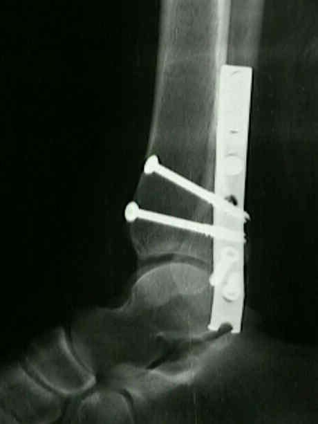
- one third tubular plate is applied to lateral (or posterolateral) fibular surface and is held w/ a bone holding clamp;
- plate is secured w/ 3.5 mm cortex screws proximally and 4.0 mm cancellous screws distally;
- it is usually possible to place 2 or 3 screws distal to frx and 3 screws proximal to the fracture;
- distal screws should engage the medial cortex of the fibula but not protrude into the fibulotalar joint;
- there is no reason, however, to grossly undersize these screws;
- wound closure:
- attempt to cover plate w/ periosteum and/or the superficial fascia;
- take care not to pass sutures into the peroneal tendon sheath since this will lead to postoperative peroneal tendinitis
The Dorsal Antiglide Plate in the Treatment of Danis-Weber Type-B Fractures of the Distal Fibula.
Rush rods versus plate osteosyntheses for unstable ankle fractures in the elderly.
 ***
***