

 - Discussion:
- tripled layered arrangement on the lateral aspect of knee;
- posterolateral part of capsule is divided into two laminae that encompass LCL, fabellofibullar, & arcuate ligaments;
- Layer I
- superficial Layer (Layer I): 2 parts;
- iliotibial tract (and its expansion anteriorly)
- superficial portion of biceps and its expansion posteriorly;
- layer I is most robust where its layers are verticle
- peroneal nerve lies on deep side of Layer I, just posterior to biceps femoris;
- Layer II
- layer II of lateral side includes LCL, which originates on lateral epicondyle of femur just anterior to origin of the gastrocnemius;
- its insertion on the head of fibula is continguous w/ biceps tendon;
- it is relaxed in flexion, but is probably major varus stabilizer of knee in extension;
- layers II and III are adherent to each other in a vertical line at lateral margin of the patella.
- upper fibers of patellofemoral ligaments attach to overlying iliotibial tract occur just below termination of lateral intermuscular septum at the lateral femoral epicondyle;
- anteriorly:
- is formed by retinaculum of quads, most of which descends anterolaterally and adjacent to the patella;
- posteriorly:
- is incomplete and is represented by two patellofemoral ligaments;
- proximal ligament joins terminal fibers of lateral IM septum;
- distal ligament ends posteriorly at fabella or at insertions of posterolateral patellmeniscal ligament also part of Layer II;
- it travels obliquely from patella, attaches to margin of lateral meniscus, & terminates inferiorly on lateral tibial tubercle of Gerdy (deep to the iliotibial tract);
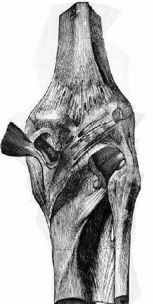
- Layer III
- deepest layer, is the lateral part of the joint capsule;
- it is attached to edges of tibia & femur circumferentially in horizontal planes at the proximal and distal ends of knee joint;
- capsular attachment to outer edge of lateral meniscus is called coronary ligament;
- popliteus tendon passes thru a hiatus in coronary ligament to attaach to femur; bare area of meniscus occurs at hiatus;
- posteriorly:
- behind overlying iliotibial tract, capsule divides into two laminae;
- superficial laminae encompasses LCL and ends posteriorly at fabellofibular ligament;
- deeper laminae passes along edge of lateral meniscus, forming coronary ligament & hiatus for passage of poplliteus tendon;
- fabellofibular ligament alone reinforces capsule in 20% of pts;
- both arcuate ligament & fabellofibular ligament reinforce posterolateral aspect of the capsule in 2/3 of pts;
- when fabella is large, arcuate ligament is not present & fabello- fibular ligament is strong;
- fabellofibular ligament will be found deep between biceps tendon and lateral head of gastrocnemius;
- may be as large as LCL;
- fabella:
- when fabella (cartilaginous fabella) is absent, fabellofibular ligament is also absent and only arcuate ligament is present;
- when cartilagenous fabella is present, fabellofibular ligament & arcuate ligament will be present
Anatomy of the junction of the vastus lateralis tendon and the patella.
Knee dysfunction secondary to dislocation of the fabella.
The incidence of fabellae in osteoarthrosis of the knee.
Classification of knee ligament instabilities. Part II. The lateral compartment.
Year Book: Lateral Ligament Injury of the Knee.
The structure of the posterolateral aspect of the knee.