

- Discussion:
- medial and lateral muscle bellies each carry long flaps of skin and fascia from the posterior calf and are most often
used as myocutaneous flaps; these flaps are most useful for upper tibial defect reconstruction;
- Contra-indications:
- knee dislocation or supracondylar fracture which may injure the sural arteries;
- in these cases consider arteriogram;
- Blood Supply:
- each head of gastrocnemius is supplied by the sural artery, which is branch of the popliteal artery;
- sural artery passes directly into proximal portion of both heads, (about 3 cm above the joint line) where it
arborizes and then travels longitudinally along length of muscle;
- there are also direct fasciocutaneous arteries arising from popliteal artery as well as septocutaneous vessels
arising from trifurcation of the popliteal artery of the leg;
- Medial Head:
- for defects over knee, medial head of gastrocnemius covers knee joint (& patella, if origin from medial femoral condyle is divided);
- medial head of the gastrocnemius is ideally suited to cover exposed bone over the proximal 1/3 of the tibia;
- medial or lateral heads of the gastrocnemius muscle can be expended with little or no deficit when walking or in normal running;
- amoung disadvantages of the muscle flap is that a portion of proximal achilles tendon must be divided to free medial
or lateral head of gastroc muscle;
- Surgical Approach for Medial Gastroc Flap:
- longitudinal incision which parallels the posterior border of the fibula, from the tibial plateau to a point 10 cm above the ankle;
- if flap is to be tunneled under a skin bridge, the incision should be placed even more posteriorly, to ensure adequate skin bridge;
- saphenous vein is left intact;
- gastrocnemius is separated from the overlying subQ tissue;
- avascular plane is developed between the medial head of the gastroc and soleus;
- at the median raphe, small vessels may be identified between the gastroc and soleus;
- median raphe:
- at times it is difficult to identify the midline raphe between medial and lateral head of the gastroc;
- gastrocnemius muscle is composed of two heads that are paritially fused in the midline of the calf, but more
proximally the two heads can be bluntly separated (at the popliteal fossa);
- proximally sural nerve traverses the midline between the two heads, but then passes lateral to the raphe;
- distally the attachment of the medial head is released w/ a small portion of of the achilles tendon attached to the flap;
- after its attachment to Achilles tendon is freed, the neurovascular pedicle is isolated;
- fascia on the deep surface of the muscle can be scored longitudinally to increase the breadth of the flap;
- if indicated, the pedicle can be dissected to its origin and muscle's femoral attachments divided, w/ care to avoid sural artery damage;
- Lateral Head:
- allows coverage of exposed lateral aspect of knee joint to the patella;
- it is shorter in length;
- during dissection of this flap, peroneal nerve is at risk;
- it is separated from tibia by fibula, lateral compartment, and anterior compartment;
- longitudinal incision is made 3 cm posterior to fibula;
- peroneal nerve is identified at the neck of the fibula


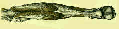
The gastrocnemius myocutaneous flap used as a over for the exposed knee prosthesis.
Salvage of jeopardized total-knee prosthesis: the role of the gastrocnemius muscle flap.
Making the most of the gastrocnemius muscles.
The medial gastrocnemius myocutaneous flap.
Year Book : Why the Denervated Gastrocnemius Muscle Flap Should Be Encouraged.
Aesthetic considerations of the medial gastrocnemius myocutaneous flap.