- See:
- Distal Radius Frx Menu
- External Fixators for Distal Radius Frx
- Intra-Articular Fractures of the Distal Radius
- Unstable Distal Radius Frx
- Discussion:
- clinical outcome studies
- Radiographs:
- r/o concomitant scapholunate dissociation;
- Anatomic Considerations:
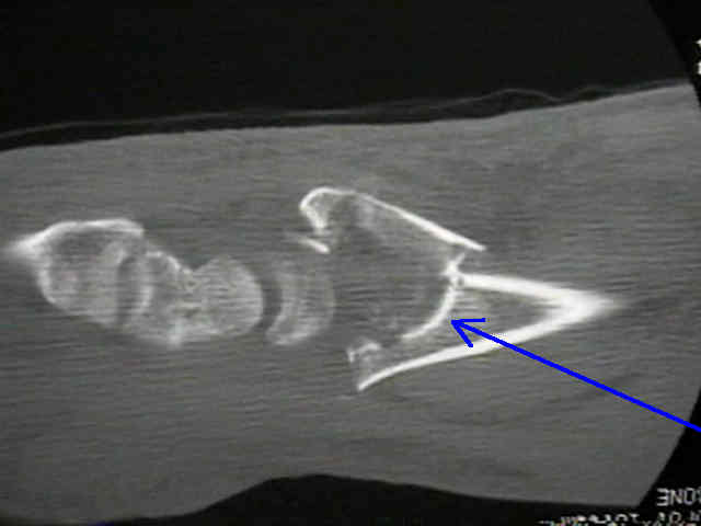
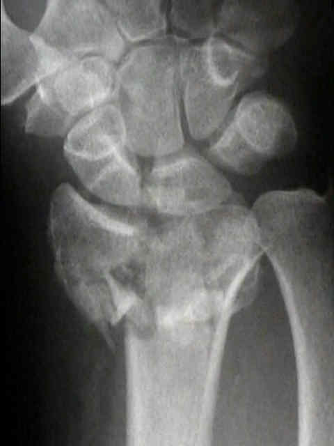
- die punch fragment:
- following fixation of the radial styloid fragment, the remaining depressed articular fragments are elevated and reduced;
- reduction is facilitated w/ traction, direct pressure, or with use of a small incision and application of pointed reduction clamps;
- if reduction can not be performed closed, then a limited open reduction can be performed;
- wires can be inserted transverse across the subchondral portion of the distal radial articlular surface, either through the ulna
and across the RU joint, or directed transversely thru the radial cortex to lie just under the subchondral surface;
- metaphyseal comminution:
- w/ high energy frxs or w/ metaphyseal comminution, consider combination of external fixation and bone grafting in order to
prevent late collapse of the articular reduction;
- Radiographs: - look for concomitant scapholunate dissociation;
- Specific Techniques:
- it is essential to hold the fracture closed reduced as possible while the pins are inserted inorder that there is minimal skin traction
against the pins;
- consider positioning distal forearm on a stack of towels which allows wrist be maximally palmar flexed which helps w/ reduction,
which facilitates pin insertion (hand and thumb are moved out of way), and which allows easy flouroscopy since distal
forearm rests parallel to ground on towels;
- references:
- Intraarticular fractures of the distal radius: a cadaveric study to determine if ligamentotaxis restores radiopalmar tilt.
- Severe fractures of the distal radius: effect of amount and duration of external fixator distraction on outcome.
- Biomechanical analysis of pin placement and pin size for external fixation of distal radius fractures.
- Supplemental pinning improves the stability of external fixation in distal radius fractures during simulated finger and forearm motion..
- A biomechanical comparison of different wrist external fixators with and without K-wire augmentation
- Percutaneous wire fixation of distal radial fractures: is it preferable to bury the wires?
- Percutaneous fixation with K wires vs volar locking-plate fixation in adults with dorsally displaced fracture of
distal radius: five-year follow-up of a randomized controlled trial
- extra-focal pinning techniques:


- kapandji's technique:
- references:
- Two Procedures for Kirschner Wire Osteosynthesis of Distal Radial Fractures. A randomized trial.
- Intrafocal (Kapandji) pinning of distal radius fractures with and without external fixation.
- T-pin - Union Surgical:
- dorsal pin placement:
- single dorsal transfixion K-wire yields the greatest reduction in fragment motion in the flexion/extension plane;
- single 0.065-inch (1.6 mm) K-wire is used to augment fixation;
- wire is drilled at a 45° angle in sagittal plane from dorsal lip of distal radius, across osteotomy site and through volar
cortex (dorsal transfixion wire);
- starting point is positioned just distal to Lister's tubercle;
- ref: Dorsal pin placement and external fixation for correction of dorsal tilt in fractures of distal radius.
- trans-ulnar technique:
- ulnar-radial pinning with fixation of the DRUJ
- K wires are placed thru distal ulna into the reduced distal radius;
- technique avoid dorsal sensory branch of radial nerve;
- there is enhance stability with this technique since there is bicortical pin placement thru the ulna;
- disadvantage: need to immobilize R-U joint w/ long arm cast;
- references:
- Comminuted fractures of the distal end of the radius treated by ulnar pinning
- The history and evolution of percutaneous pinning of displaced distal radius fractures.
- Trans-ulnar percutaneous pinning of displaced distal radius fractures: a preliminary report.


- Bone Grafting: (see bone graft harvest techniques and bone graft menu);
- elevation of impacted fracture fragments often results in a metaphyseal fracture defect;
- bone grafting via a limited incision over the frx site fills in the fracture site defect and helps prevent fracture collapse;
- the bone graft attempts to hold the reduction in place, and in a sense helps take the place of external fixation;
- it is often easier to perform percutaneous pinning and bone grafting at 10 days from injury since this allows the frx site to
become slightly sticky;
- reference:
- Augmentation of distal radius fracture fixation with coralline hydroxyapatite bone graft substitute.
- Assessment of Reduction: Is external fixation necessary?
- in an unstable distal radius frx w/ inadquate reduction consider the addition of external fixation + K wires;
- as noted in the study by Trumble, et al 1998, external fixation provided clear advantages in specific situations;
- in older patients, pain relief, grip strength, and ROM were significantly better when external fixation was used;
- in younger patients, external fixation provided consistently better results when there was comminution in 2 or more cortices
or when there was comminution of one surface which was greater than 50% of the metaphyseal diameter;
- restoration of radial length was more important than dorsal tilt or radial tilt, and EF afforded better restoration of length than
pinning and casting;
- ref: Intrafocal (Kapandji) pinning of distal radius fractures with and without external fixation
- Complications:
- RSD may result from pin injury to the superficial radial sensory nerve;
- RSD is avoided by making small incisions over the pin insertion site and by spreading with a hemostat;
- maintain the reduction while the surgeon uses a soft tissue protector to prevent the radial nerve from winding around the pin;
- if an assistant is not available consider applying a lubricant (K-Y) to the pin;
- references:
- Complications of distal radial fractures: pins and plaster treatment.
- The thermal effects of skeletal fixation-pin insertion in bone.
- References:
Unstable distal radius fractures treated by modified Kirschner wire pinning: anatomic considerations, technique, and results.
Percutaneous and limited open reduction of the articular surface of the distal radius.
Treatment of comminuted distal radius fractures with pins and plaster.
Percutaneous Pinning of Distal Radius Fractures: A Biomechanical Study.
Percutaneous wire fixation of distal radial fractures: is it preferable to bury the wires?
Percutaneous pinning of distal radius fractures restores motion over long-term. The minimally invasive technique bucks the popular trend toward ORIF.
Study finds plating distal radius fractures effective but costly
Good long-term results, few complications seen with pinning distal radius fractures
Stable distal radius fracture fixation possible with threaded pinning method
T-Pin Distal Radius Fixation System
Ulnocarpal stabilization in the management of comminuted fractures distal end radius
Percutaneous cannulated screw fixation in the treatment of distal radius fractures.
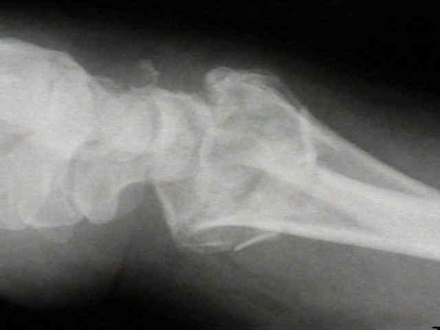
- Case Examples:


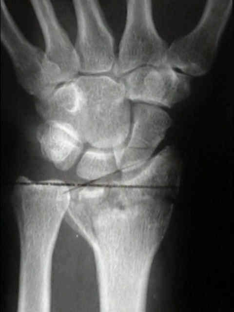
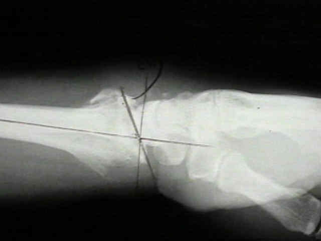
- this 40-year-old patient underwent application of a external fixator prior to insertion of pins;
- note that prior to pin insertion, the external fixator was distracted to help maintain the reduction, but at the end of the case, the distraction was released and the reduction was rechecked w/ flouro;
- it is essential that the MP joints achieve full flexion at the end of the case, as overdistraction can lead to MP join clawing