

- See: Anatomy of Carpal Tunnel
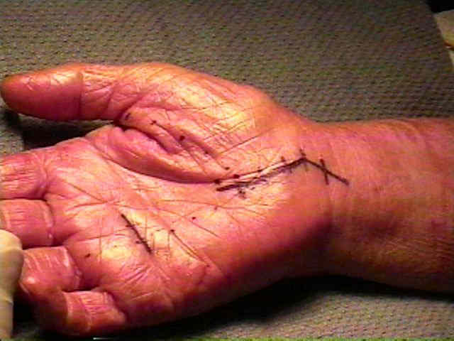
- TCL Incision:
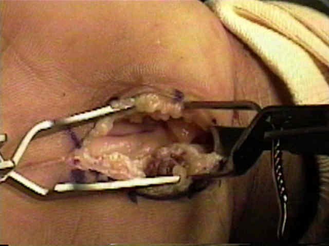
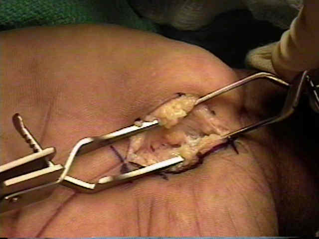
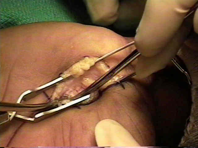
- transverse carpal lig is exposed, but this is generally obscured by origin of thenar & hypothenar muscles, which come to lie in opposition in midline;
- palmar fascia is incised, & nerve is identified just proximal to the carpal canal;
- this is carried out with either a proximal extension of the incision or with a separate twin incision technique;
- blunt dissection is used to identify the distal edge of the TLC;
- first identify the superficial palmar arch, which lies distal to the distal edge of the TLC;
- realize that the distal fibers of the TLC extend distal to the scaphoid and hook of the hamate, and that the distal free edge is best identified near the midline between the thenar and the hypothenar muscles groups;
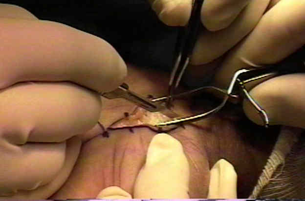
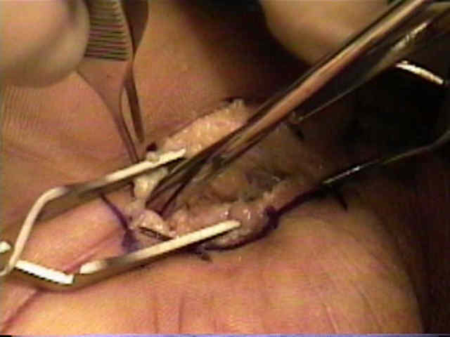
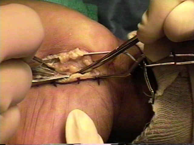
- a Crile clamp is used to identify the distal opening of carpal tunnel and then to elevate it which allows it to be incised from distal to proximal (while very carefully observing that the median nerve is not injured);
- guide is passed proximally under retinaculum so that incision in it can be continued proximally;
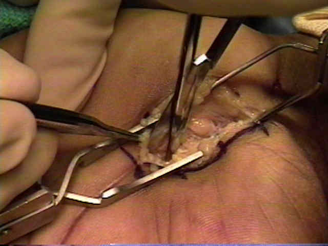
- some note that an anomolous motor branch might be injured when instruments are placed blindly into carpal tunnel;
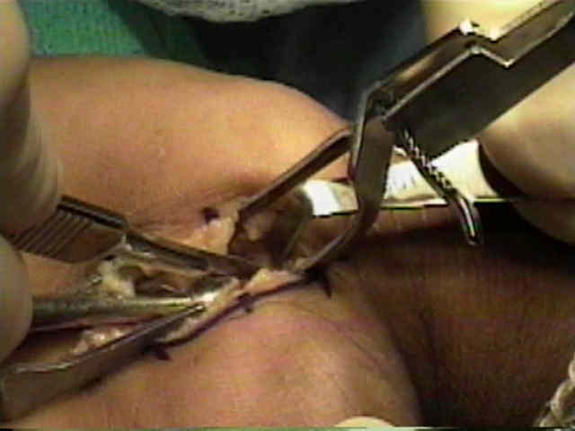
- incision must be kept to ulnar side of median nerve & palmaris longus (if present) to avoid damaging the motor branch of the nerve;
- special care must be taken to avoid the motor branch of median nerve which comes off in a radial palmar direction near the terminal arboziation of the nerve;
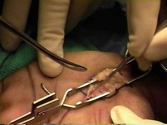
- by incising on the ulnar side of the carpal tunnel, the TLC will remain ulnar to the median nerve, which helps prevent bowstringing;
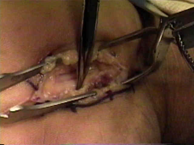
- w/ ulnar dissection, need to identify and avoid superficial palmar arch;
- note that the deep motor branch of the ulnar nerve courses around the hook of hamate and can be injured;
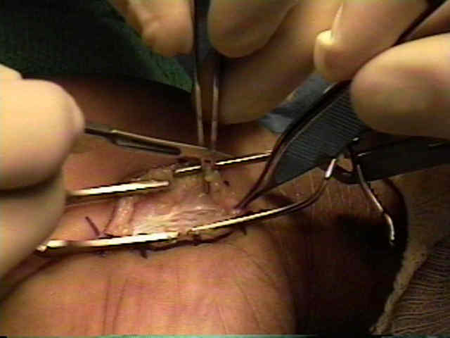
- incising ligament over an instrument placed in the carpal canal helps to avoid damaging the median nerve;
- dissection is extended proximally until deep fascia of forearm is reached;
- alternatively, retinaculum can be incised from its proximal edge after carefully identifying median nerve & keeping to its ulnar side;
- one should investigate whether motor branch is compressed by a fascial ring that surrounds the nerve as it enters the thumb muscles
Anatomy of the flexor retinaculum.
Anatomic relations between the median nerve and flexor tendons in the carpal tunnel: MR evaluation in normal volunteers.
Variations of the median nerve in the carpal canal.