- Discussion:
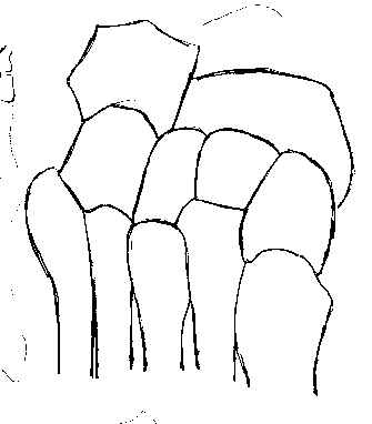
- anatomy of the midfoot:
- many orthopaedists have moved away from pin fixation because fixation must be rigid enough to prevent transverse plane & dorsoplantar motion of TMT joint and be maintained for at least 12-16 weeks;
- this exceeds the length of time that pins can be left in place;
- an alternative, is percutaneous insertion of cannulated screws, which allows rigid fixation, w/o risking wound slough which may occur following open reduction;
- Radiographs:
- w/ questionable injury, consider wt bearing AP view to assess 1-2 interval;
- if standing AP is unacceptable to the patient then consider CT scan;
- Fixation:
- technique of reduction:
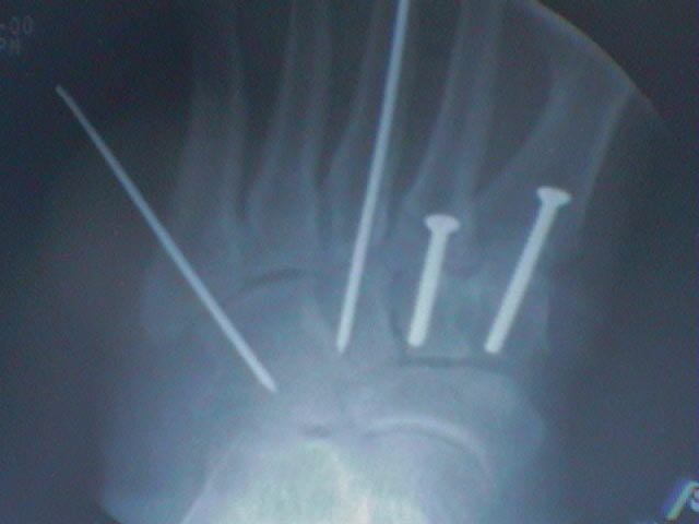
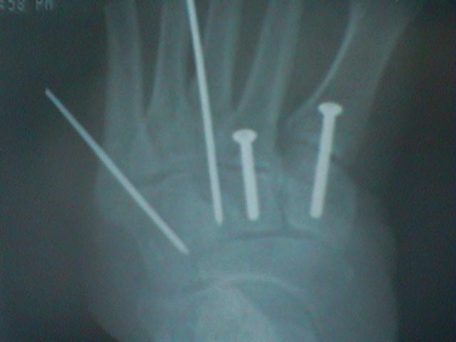
- fixation is performed w/ 0.062 inch K wires;
- isolated dislocation of 1st metatarsal:
- consider placement of 2 K wires thru the shaft into the cuneiforms;
- homolateral and divergent dislocations:
- insert one K wire medially thru 1st metatarsal into cuneiform;
- insert one K wire laterally thru 5th metatrsal into cunboid or into the calcaneus;
- non wt bearing cast is worn for 6 weeks;
- as an option, cannulated screws can be placed over the K wires, for more reliable fixation;
- Case Examples:
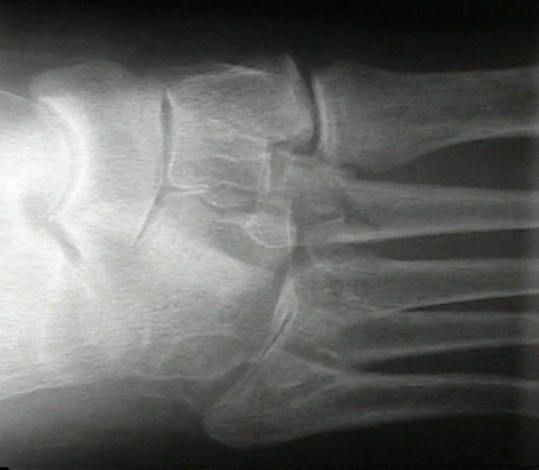
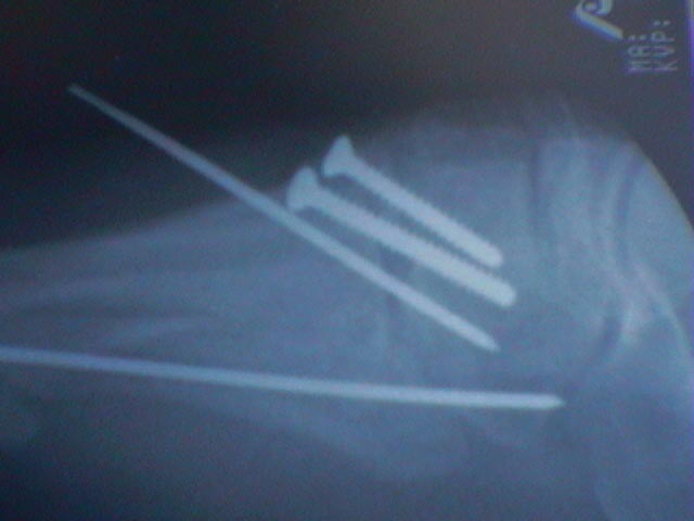
- 25 year old athelete who was injured during a pile up on the field;
- standard radiographs were interpreted as normal, but this wt bearing film clearly shows the injury;
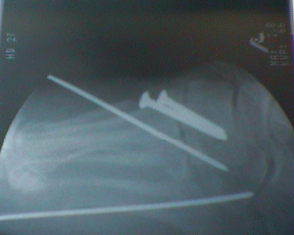
- small incisions were made over the base of the second metatarsal and medial cuneiform;
- a large tenaculum clamp was used to oppose the base of the second metatarsal to the cuneiform;
- after adequacy of the reduction had been confirmed, a cannulated screw was inserted