- See: STSG
- Discussion:
- full thickness grafts transfer all organs contained in the dermis in addition to some Pacinian corpuscles (when the graft is taken from the
palm or sole);
- these grafts allow more durable coverage and more sensation than STSG;
- donor site:
- graft should be harvested from a hairless areas where the skin is redundant;
- most common sites are volar wrist crease, volar elbow crese, and inguinal region (or in the area between the abdomen and the tigh),
and from the ulnar side of the hypothenar eminence;
- grafts need to be elliptical with the longitudinal axis in langer's line;
- beaware of pigmentation differences between donor and recipient site;
- FTG applied to the hand:
- consider the instep of the sole, which which transfer skin w/ Pacinian and Meissner's corpuscles, as well as matching color;
- ulnar side of the hypothenar eminences provides glabrous skin from site that is readily available and can be closed primarily,
resulting in an inconspicuous scar;
- Technique:
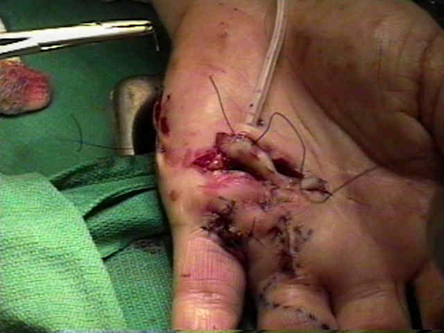
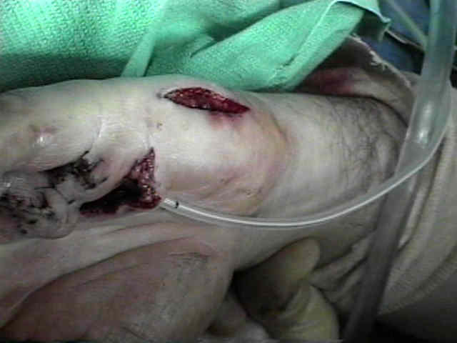
- wound debridement:
- the recipient site for a full thickness skin graft will contract after being excised;
- ie, the skin edges around around the margin of defect retract, as soon as the opposing skin tension is gone;
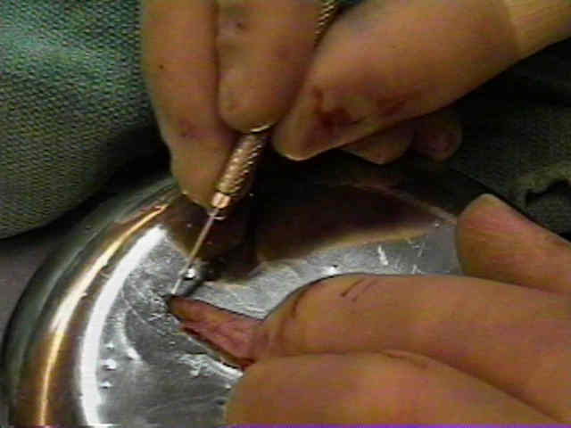
- graft harvest:
- outline an oval paddle of skin from appropriate donor site;
- ensure that outlined graft is of appropriate size;
- the defect can be outlined w/ sterile paper and then applied to the donor site;
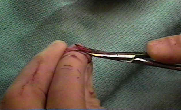
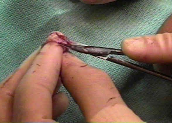
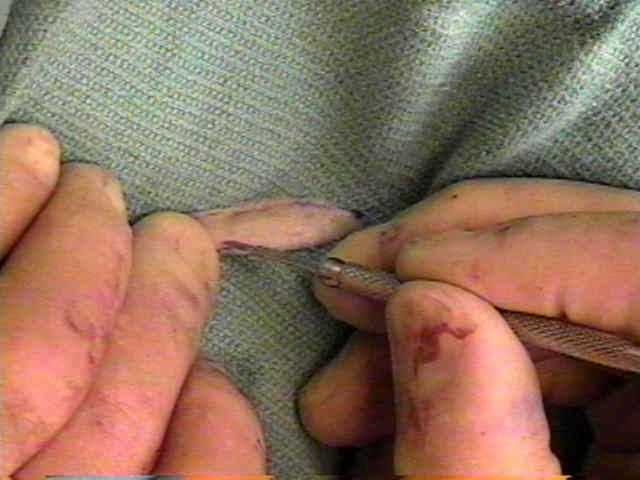
- place tension on the skin incision which facilitates separation of the subdermal fascia from the skin;
- excise the subdermal fascia and undermine the subQ tissue inorder to facilitate wound closure;
- on the back table, place the graft under tension, and remove the remaining subQ tissue;
- pie crust the graft w/ a scapel, to allow egress of fluid;
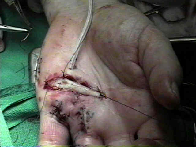
- Graft Application and Postoperative Care:
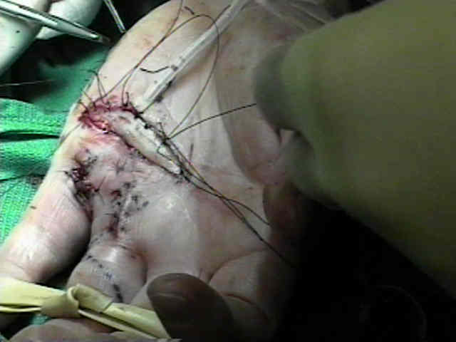
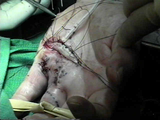
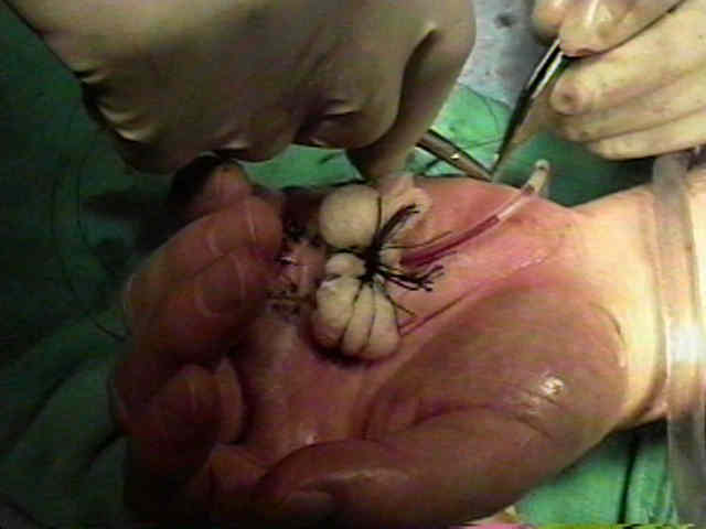
- in most cases the graft will be tied down at the edges w/ a non absorbable suture, and then the suture limbs will be used to secure the
dressing;
- the dressing consists of nonadherent gauze (adaptic) following by gauze soaked in Bunnel's solution, followed by more Adaptic;
- the limb is then splinted to reduce motion;
- note that at the initial dressing change, the graft may appear non viable due to sloughing of the stratum corneum;
- the underlying dermis, however, continues to be viable