

- Radiographs: 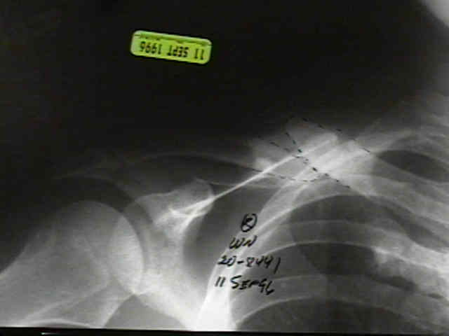

- Positioning:
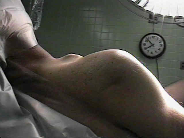
- supine, HOB elevated to 30-40 deg;
- head is held with a dedicated ring holding device (McConnel);
- one rolled towel is placed underneath the operative shoulder and one rolled towel is placed around the contralateral hip / rib cage;
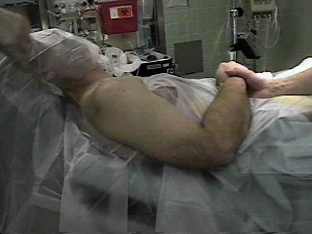
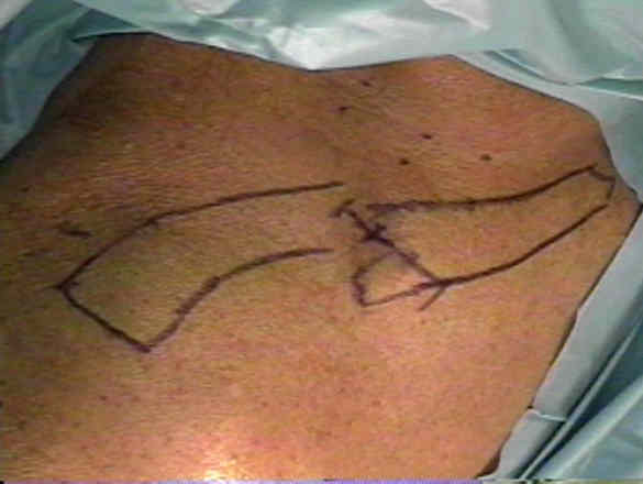
- surgical draping should allow exposure from SC joint to the posterolateral aspect of the shoulder;
- surgical draping should not interfere w/ taking intra-operative radiographs (consider placing the radiographic cassette under the shoulder prior to draping);
- in cases of non-union, the iliac crest is also prepped out for bone grafting;
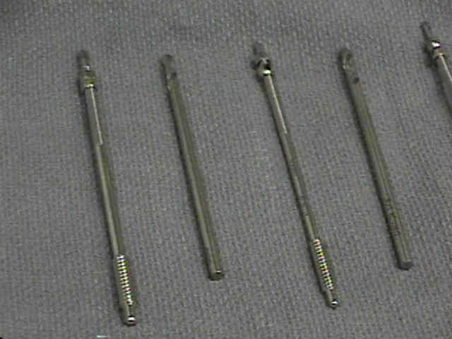
- Implants:
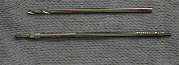
- modified Hagie pins have a blunt tipped end w/ large threads and a fine tipped end which can accomodate a hexagonal compression nut;
- the blunt end with large threads is used to secure the medial fragment (and prevent migration);
- pins come in several sizes, each w/ an appropriate sized drill;
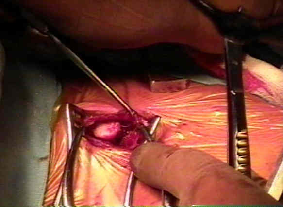
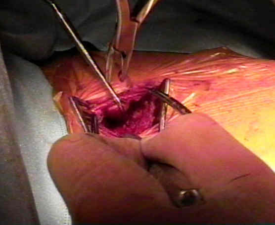
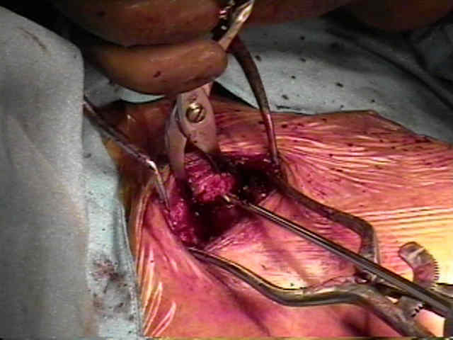
- Fracture Site Debridement:
- in cases of non-union, fibrous tissue is debrided from the fracture site, until both bone edges are bleeding;
- in cases of malunion, callus is debrided until the normal anatomy is restored;
- each bone end should be feathered with an osteotome, and following screw insertion, bone graft will be applied over the fracture site;
- periosteum is stripped on either side of the fracture site;
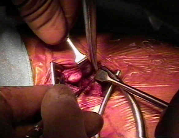
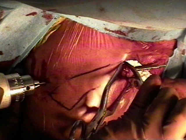
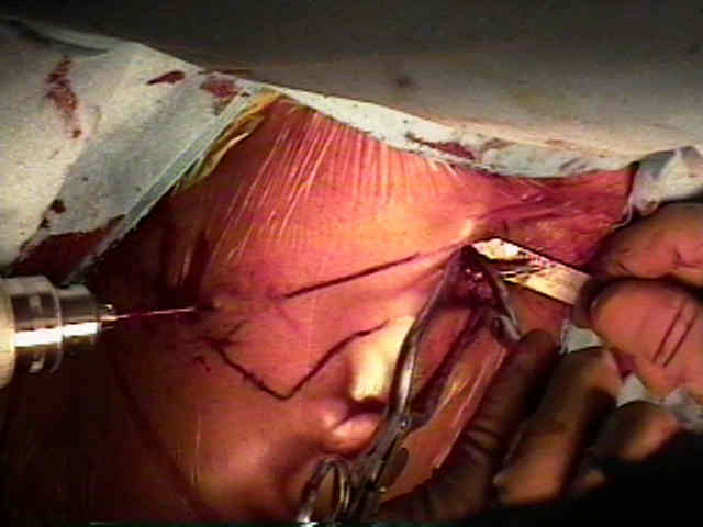
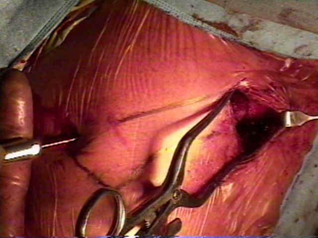
- Frx Reduction:
- have 2 "lobster claw" type reduction clamps available inorder to manipulate the frx ends;
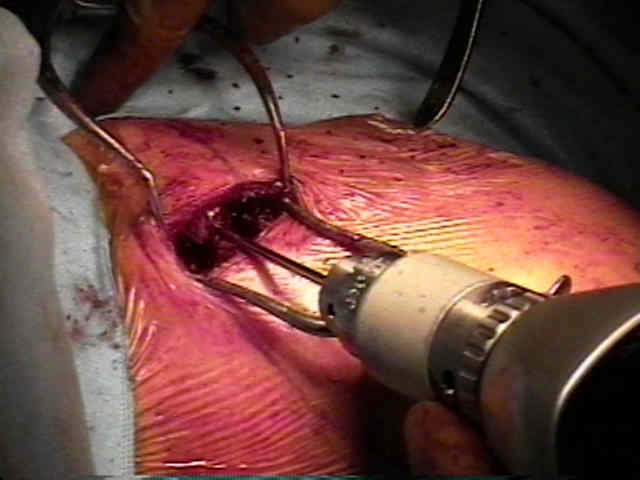
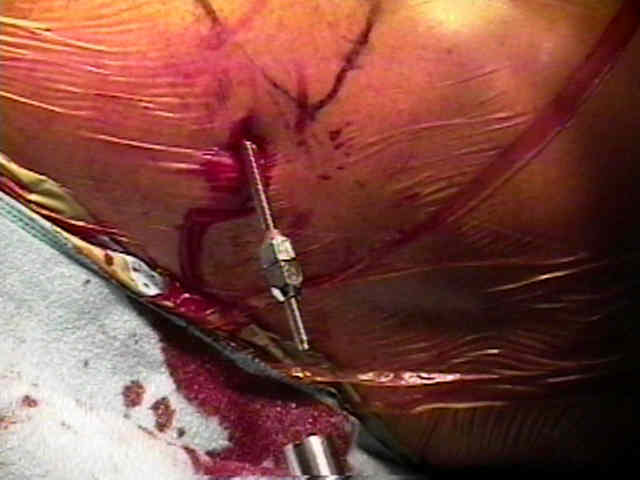
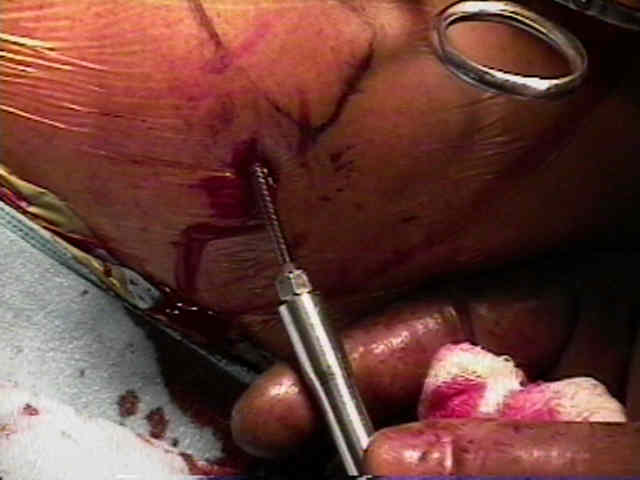
- Reaming of Medial Fragment:
- the intramedullary canal of the medial fragment is reamed w/ a hand drill;
- take care not to perforate the anterior cortex;
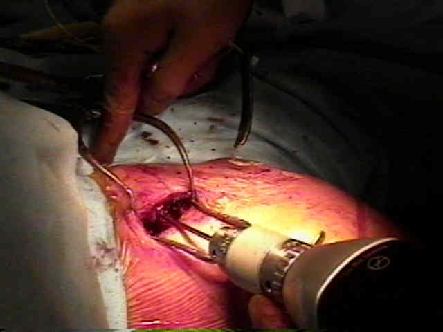
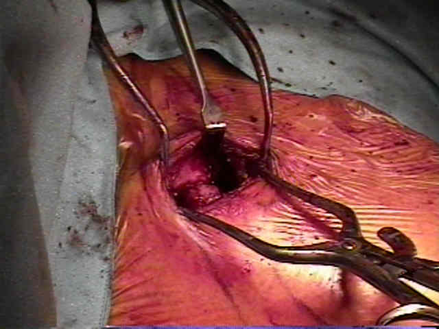
- Retro-grade Screw Insertion: 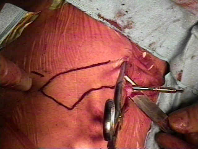
- Pin is Pulled Back to Osteotomy Site:
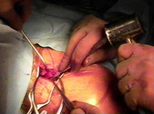
- Antegrade Insertion by Hand:
- prior to this step, the reduction clamps are re-applied across the fracture site;
- two hexagonal nuts are applied to the lateral end of the Hagie pin (the adjacent nuts will not migrate), and subsequently a hexagonal screw driver is applied and is used to drive the pin anteriorly across the fracture site;
- once the medial threads have passed the fracture site, fracture compression usually takes place;
- remove one of the two hexagonal nuts, and then advance the remaining screw up to the fracture site, in order to apply compression;
- Application of Hex Nut Compression
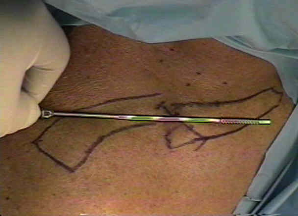
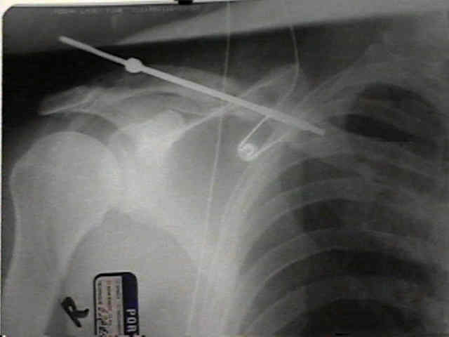
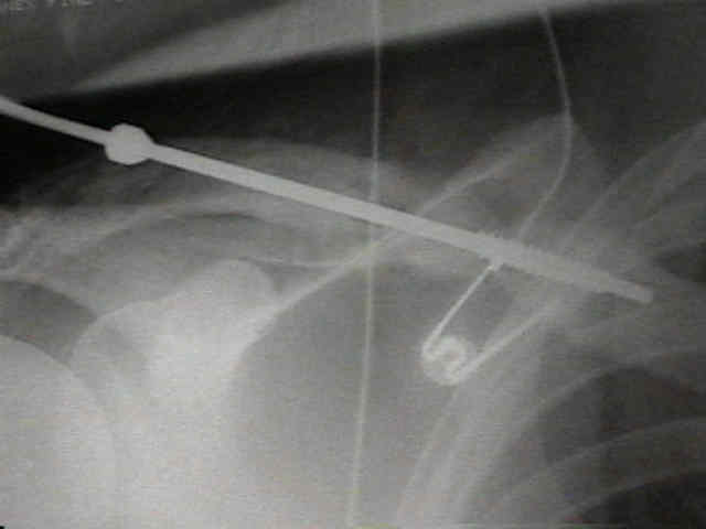
- Intra-Op Radiographs:
- Pin Clipped Beneath Skin:
- note that conventional pin cutters will leave the IM pin too prominent beneath the skin, and this may lead to pin erosion through the skin during the first several weeks of the post op period;
- consider clipping the pin beneath the skin, and then back the pin out of the skin with a drill a distance of 1.5 cm;
- then reclip the pin beneath the skin with the pin cutter;
- reattach the drill to the pin and drive the pin medially about 1.5 cm;
- the pin should now be well below the level of the subcutaneous tissue and will not hurt the patient during activies of daily living;
- Post Operative Care:
- in order to reduce rotatory forces, patients are discouraged from raising the operative arm above the horizontal;
- the pin is removed at 6 weeks for acute fractures;
- w/ non-union, the pin is removed when there is radiographic signs of healing (usually 22 weeks);
- technical tip: consider using power driver with Jacobs chuck (reverse mode) for easy pin removal;
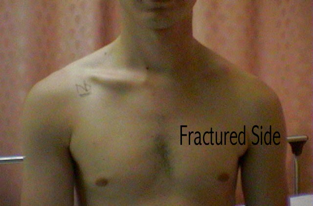
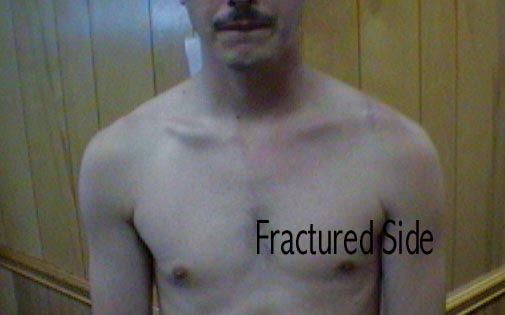
- outcome: (before and after photos)
- Complications:
- brachial plexus palsy:
- may be associated with use of beach chair positioner and aggressive mobilization of fracture ends as they are delivered out of the incision, just prior to fracture site reaming
Nursing Checklist:
The treatment of nonunion fractures of the midshaft of the clavicle with an intramedullary Hagie pin and autogenous bone graft.
Brachial Plexus Palsy After Intramedullary Fixation of a Clavicular Fracture. A Report of Three Cases.
Complications of clavicle fractures treated with intramedullary fixation
Midshaft clavicular fractures: comparison of intramedullary pin and plate fixation
- Implant:
- the IM Clavicular Pin is produced by Depuy Orthopaedics