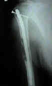
- See: Humeral Frx - Main Menu
- humeral shaft fractures
- Retrograde IM Nailing:
- main advantage over antegrade nailing, is that violation of the rotator cuff is not necessary (and therefore shoulder motion is not affected);
- retrograde nailing is also preferred over antegrade nailing for distal shaft fractures;
- main disadvantage is technical difficulty and the need for prone position;
- as noted by Lin et al. (1998), for distal frx, retrograde nailing showed more initial stability and stiffness as compared to antegrade nailing (the
opposite being true for proximal frx);
- initial stability is improved by tight grip of the distal cortex on the nail at its entry point;
- positioning: prone position, w/ torsal placed near the edge of the table, and arm resting on arm board, and the elbow flexed to 90 deg (ensure
that adequate flourscopic views are possible);
- limited posterior approach is made over distal humerus, from the olecranon tip to a point 8 cm proximal;
- avoid entering the elbow joint if possible to avoid periarticular scarring;
- entry site:
- located about 2.5 cm above the olecranon fossa (the roof of the olecranon fossa is not used for entry);
- long elliptical entry portal made at the superior border of the olecranon fossa;
- this entry hole has a minimal effect on bone strength;
- make the initial entry using multiple small drill holes made perpendicular to the cortical surface;
- a rotating burr will help enter the medullary canal without trauma;
- beaware of potential fissure and/or avulsion at the entry site;
- in the study by Strothman D, et al (2000), the authors studied the loss of strength in the distal humerus that
resulted from the creation of two different entry portals;
- metaphyseal entry portal:
- created by reaming in the center of the dsital metaphyseal triangle (two cm proximal to olecranon fossa);
- reduced the torque to failure to 71 % of the intact specimen and the energy absorbed to failure to 37% of the that of the intact tissues;
- olecranon fossa entry portal:
- entry portal is created in the proximal slope of the olecranon;
- reduced torque to failure to 55 % of that of intact specimens and energy aborbed to failure to 18 % of that of the intact specimens;
- the authors recommended great caution is allowing these patients to use the upper extremities for wt bearing activies (ie bed to chair transfers);
- consider careful flourscopic imaging inorder to avoid excessive reaming of the anterior humeral cortex inorder to avoid fracture;
- IM nail diameter: standard size is 7.5 mm (smaller and larger diameter nails should be available);
- IM nail length: distal end of the nail should lie 2-3 cm above the olecranon fossa, and proximally the nail should protrude just slightly into the humeral head;
- complications:
- radial nerve palsy:
- may occur in about 4% of cases;
- references:
- Closed retrograde Hackethal nail stabilization of humeral shaft fractures.
- Retrograde locked nailing of humeral shaft fractures. A review of 39 patients.
- Biomechanical comparison of antegrade and retrograde nailing of humeral shaft fracture.
- Retrograde nailing of humeral shaft fractures: a biomechanical study of its effects on the strength of the distal humerus.
Intramedullary fixation of humeral shaft fractures.
Intramedullary fixation of complicated fractures of the humeral shaft.
Intramedullary stabilization of humeral shaft fractures in patients with multiple trauma.
Locked nailing of humeral shaft fractures. Experience in Edinburgh over a two-year period.
A biomechanical comparison of intramedullary nailing systems for the humerus.
Treatment of humeral diaphyseal fractures with Hackethal stacked nailing: a report of 33 cases.
Seidel intramedullary nailing of humeral diaphyseal fractures: a preliminary report.
Closed intramedullary fixation of humeral shaft fractures.
Distal interlocking during intramedullary nailing of the humerus.

Intramedullary stabilization of humeral shaft fractures in patients with multiple trauma.
Antegrade Interlocking Nailing of Acute Humeral Shaft Fractures.

