- See:
- Ficat Classification
- Enneking's Stages of Osteonecrosis
- Discussion:
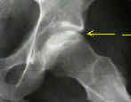
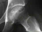
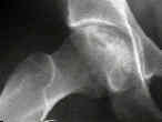
- combination of osteoclastic resorption and new bone formation in revascularized areas creates x-ray appearance of mottled density;
- when collapse of segment of necrotic bone occurs, the compression of more bone into smaller area also produces increased x-ray
density;
- there are several anatomic reasons for the increased density of necrotic bone as seen radiographs;
- if radiographic evaluation is consistent w/ diagnosis of AVN, no additional diagnostic tests are necessary;
- note however, that plain radiographs are often inadequate to make the diagnosis;
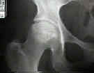
- AP View:
- usually demonstrate the principal area of involvement;
- because anterior and posterior acetabular margins overlap the superior portion of femoral head, subtle
evidence of osteosclerotic or cystic changes in subchondral regions may be missed;
- Lateral View:
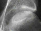
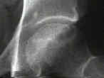
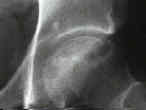
- because AP views of the hip may be difficult to interpret, it is necessary to evaluate frog-leg lateral x-rays of the femoral head;
- cross-table lateral x-ray is not satisfactory because architectural details
of the femoral head are obscured by the soft tissues that overlie this area;
- lateral radiographs also allow staging purposes since it is often anterior segment
of the femoral head that first collapses or exhibits the crescent sign


