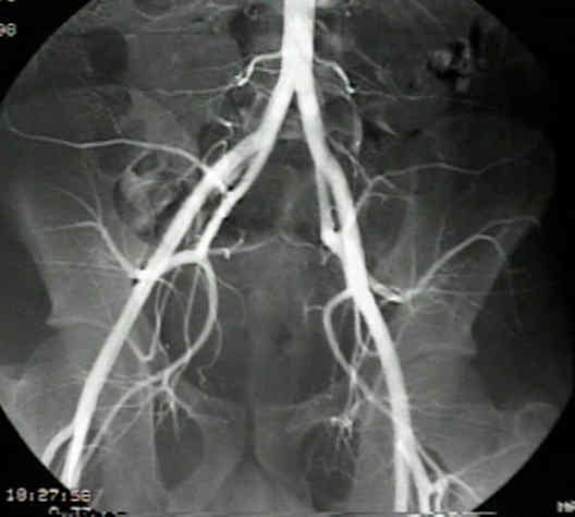
- Discussion:
- anterior approach:
- femoral artery may be palpated in femoral triangle, & may be used as a guide in aspirating the hip joint;
- palpate the femoral pulse just as it exits the inguinal ligament;
- entry point is one inch lateral to the artery (at the inguinal ligament) and one inch below the inguinal ligament;
- going lateral 1 inch will also make entry site approx 1 in below ligament;
- needle entry is then straight down into the lateral half of the joint cavity;
- disadvantages: if the surgeon is not in the capsule when the contrast dye is injected, then contrast material will
collect and will obstruct needle visualization;
- lateral approach:
- greater troch is palpated & needle inserted just anterior to its superior tip;
- needle is directed 45 deg cephalad, & parallel to table (pt is supine);
- femoral neck will usually be met & needle can then be directed sl cephalad and proximal to enter the hip joint;
- greater trochanter is palpated, & needle is inserted from side, in front of its upper margin and approx parallel to femoral neck, so that needle
enters capsule obliquely after passing thru atachments of gluteus medius & minimus;
- disadvantages: in patients with large thighs, the needle may not be long enough to reach the joint;
- medial approach:
- needle is inserted just posterior to the insertion of the adductor longus muscle, and anterior to the gracilis;
- flouroscopy is then used to direct the needle into the hip joint

