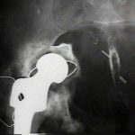
(see also: Bone Grafting for Acetabular Defects)
Discussion
- it is probably the result of remodeling of weak, medial acetabular bone after multiple, recurring stress fractures.
- intrapelvic protrussion of the acetabulum may be primary or secondary;
- protrusio acetabuli is not found only in inflammatory arthritides;
- most cases are in patients with osteoarthritis.
primary protrusio: Otto Pelvis (Arthrokatadysis)
- primary protrusio acetabuli characterized by progressive protrusio in middle aged women;
- is bilateral in 1/3 of pts & causally related to osteomalacia
- large cortical-cancellous bone grafting may be required using pt's femoral head in a primary arthroplasty as well as a large acetabular component;
- primary form, Otto pelvis (arthrokatadysis), involves both hips, occurs most often in females, & causes pain & limitation of motion at a relatively early age;
- varus deformity of femoral neck & arthritic changes are common;
secondary protrusio 
- secondary form may be caused by femoral head prosthesis, cup arthroplasty, septic arthritis, central fracture dislocation, or THR, & may be present bilaterally in paget's, marfan's, RA, AS, & osteomalacia;
- deformity may progress until femoral neck impinges on side of pelvis;
- often, because of medial migration of the femur, the sciatic near is nearer the joint than normally;
Radiographic Diagnosis
(see also preoperative radiographic evaluation)
- Kohler's line:
- relationship of femoral head to ilioishial line;
- if femoral head is medial to Kohler's line, then protrusio is present;
- may also use radiographic line from lateral border of the sciatic notch to the medial border of the obturator foramen;
- Center Edge Angle of Wiberg;
- if CE angle is greater than 35 deg, protrusio is present;
Considerations in THR
- template preoperative leg length inequality;
- realize that adaptive soft tissue changes may not allow full restoration of leg length inequality;
- in considering the best technique, it can be helpful to contrast this with the cotyloplasty technique;
- component selection:
- acetabular component design
- the peripheral fit is the key element for fixation;
- ideally component should contain a "peripheral flare" rather than a true hemisphere inorder to prevent progressive medialization of the component;
- peripherally placed screws may also prevent medialization;
- ref: Incomplete seating of press-fit porous-coated acetabular components: the fate of zone 2 lucencies.
Surgical Technique
(see also: THR surgical technique)
dislocation of hip
- in some pts such as those w/ protrusio, neck should be divided & head removed from acetabulum in a retrograde fashion rather than risk fracture;
- note that the sciatic nerve may be closer in proximity to the femoral neck than is usually seen;
reaming technique
- it is essential not to deepen the acetabulum while reaming;
- medial wall of the acetabulum is usually thin or may be partially membranous, and should not be penetrated;
- surgeon should ream inorder to obtain good peripheral fit;
- peripheral reaming technique must be exact because all residual cartilage must be removed for ingrowth, while the peripheral subchondral surface must be preserved, since this will provide the structural support for the implant;
need to establish proper offset
- failure to restore normal lateral offset may cause the greater trochanter to inpinge off of the anterior edge of the acetabulum (leading to posterior instability);
- the peripheral fit dictates the offset;
- cups with the option of acetabular screws allows the surgeon to check the depth of the cup position, can allow for more graft placement through screw holes, and scews may prevent medial cup migration;
bone grafting
(see also: bone grafting for acetabular defects)
- w/ bone grafting a noncemented cup be placed in a more lateral and anatomic position and secured with acetabular screws;
- femoral head autograft is one immediate option;
- in young pt w/ acetabular protrusion secondary to longstanding inflammatory arthritis, most authorities advise strengthening thin and medially displaced medial wall of the acetabulum w/ placement of a block cancellous autograft taken from the femoral head of the patient;
- large cortical-cancellous bone grafting may be required using pt's femoral head in a primary arthroplasty as well as large acetabular component;
outcome studies
- in the report by E. Garcia-Cimbrelo (2000):
- authors followed 148 primary THR with acetabular protrusion between 1972 and 1990;
- 62 with a mild protrusion were classified as group 1, 54 with moderate or severe protrusion as group 2 and 32 with moderate and severe protrusion which required bone grafts as group 3;
- mean follow-up was 18.3 years (3 to 24) for group 1, 17.4 years (8 to 22) for group 2 and ten years (8 to 13) for group 3.
- there were 31 revisions of the cup, 12 in group 1 and 19 in group 2;
- according to the Kaplan-Meier analysis the overall rates at 20 years were 21 ± 10.79% in group 1 and 37 ± 11.90% in group 2;
- there were 43 radiological loosenings: 22 in group 1, 21 in group 2 and none so far in group 3, at ten years;
- overall loosening rates at 20 years were 42 ± 14.76% in group 1 and 49 ± 19.50% in group 2;
- grafts were well incorporated in all group-3 hips, and the bone structure appeared normal after one year;
- the distance between the centre of the head of the femoral prosthesis and the approximate true centre of the femoral head was less in group 3 than in groups 1 and 2 (p < 0.01);
- better results were obtained in moderate and severe protrusions reconstructed with bone grafting than in hips with mild protrusion which were not grafted.
- weakness of this study is that the authors were including patients back from the 1970's and 1980's that had insertion of early press fit designs;
- authors did not specify how many cups contained a peripheral flare and how many had screw augmentation;
- ref: Loosening of the cup after low-friction arthroplasty in patients with acetabular protrusion. The Importance of the position of the cup.
References
- Radiographic measurements in protrusio acetabuli.
- Bone-grafting in total hip replacement for acetabular protrusion.
- A technique for removing an intrapelvic acetabular cup.
- Bone-grafting in total hip arthroplasty for protrusio acetabuli. A follow-up note.
- Intrapelvic migration of total hip prostheses. Operative treatment
- Protrusio Acetabuli: Diagnosis and Treatment.
- Total Hip Arthroplasty in Acetabular Protrusio
- Acetabular Protrusio: A Problem in Depth
- Surgical Technique: A Cup-in-Cup Technique to Restore Offset in Severe Protrusio Acetabular Defects

