

V. Axial Skeleton Injuries in an Austere Environment (continued)
CPT Jeanne Patzkowski, M.D.
CPT Chad Krueger, M.D.
A. Describe the initial management, resuscitation, and stabilization of pelvic fractures
IV. Stabilization
A. Provisional
1. may be performed at echelon II or III
2. sheet/binder
a. easy to use, available, provides circumferential control
b. may cause skin necrosis, nerve injury, and abdominal or extremity compartment syndromes
c. limit access to soft tissue
d. sheet technique
i. wrap longitudinally folded bedsheet circumferentially around pelvis
ii. place sheet between iliac crests and greater trochanters
iii. secure anteriorly with towel clamps
e. binder (see figure 6. Pelvic binder clinical photo; figure 9. AP pelvis with binder in proper position; figure 10. Binder improperly placed over abdomen; figure 11. APC pelvic fracture before binder placement; figure 12. AP compression pelvic fracture after binder placement.)
.jpg)
.jpg)
.jpg)
.jpg)
i. center binder over greater trochanters
ii. do not place over abdomen
(A). will not stabilize pelvis
(B). may contribute to abdominal compartment syndrome
iii. remove once hemodynamic stability achieved and vital signs unchanged with removal
3. external fixator (see figure 13. Pelvic external fixator radiograph; figure 14. Pelvic external fixator clinical photo.)
.jpg)
a. can be placed quickly and without C-arm
b. no skin necrosis or limited access to soft tissue if pins and bar carefully placed
i. do not limit access to abdomen
ii. allow space for abdominal expansion
c. only provides anterior stabilization
i. cannot stabilize posterior elements
ii. no benefit with LC-III or VS fractures
d. technique – place pins into iliac crests or supra-acetabular region
i. slight biomechanical advantage to supra-acetabular placement, but no survival benefit
ii. ensure clamps and bars do not rest against abdomen or flanks and allow for possibility of large abdominal swelling
4. C-clamp
a. may be used for posterior stabilization
b. C-arm support preferred
c. requires specific training in technique
d. may result in fracture displacement, pin site infection, cortical perforation, and nerve injury
e. technique – two pins applied to the ilium in the region of the SI joints, clamp applied anteriorly
.jpg)
1. typically at echelon V
2. open reduction internal fixation
3. percutaneous screw fixation
A. Prophylactic broad spectrum IV antibiotics – consider gram negative coverage if hollow viscus penetrated
B. Serial and extensive irrigation and debridement
C. External fixation with wound packing
D. Foreign body removal
E. Early fecal diversion with distal rectal washout
1. colostomy
2. ileostomy
3. significantly decreases incidence of infection and mortality in open pelvic fractures
F. Suprapubic cystostomy for urethral injury
VI. Associated Injuries
A. Genitourinary injuries5
1. incidence of close to 5% with pelvic fractures
a. more likely with males than females
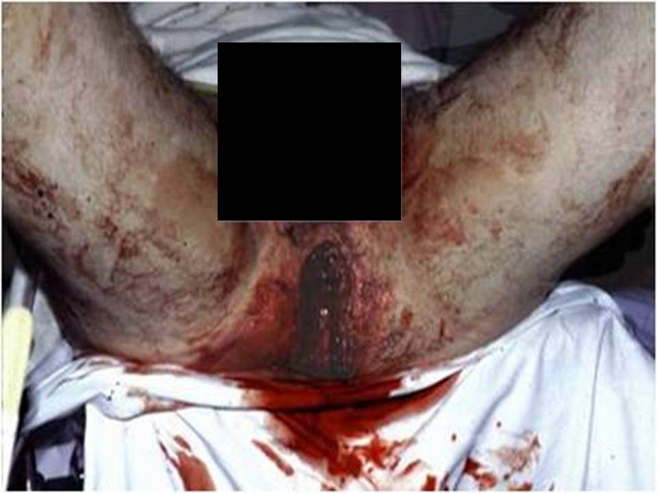
b. incidence increases with increasing severity of pelvis fracture (especially Malgaigne fractures – Figure 17)
.jpg)
2. patients with GU injuries have higher mortality rate than those without
3. symphyseal injuries associated with bladder injury, pubic rami associated with urethral injuries
B. Head injury
1. especially with lateral compression injuries of the pelvis
C. Spine injury
1. higher suspicion with vertical shear fracture patterns
References
1. Baker SP, O’Neill B, Haddon W, Long WB. The Injury Severity Score: a method for describing patients with multiple injuries and evaluating emergency care. Trauma. 1974;14(3):187-196.
2. Schulman, JE, et al. Pelvic Ring Fractures Are an Independent Risk Factor for Death After Blunt Trauma. The Journal of Trauma: Injury, Infection, and Critical Care, 2010. 68(4): p. 930-934.
3. Arthurs Z, Kjorstad R, Mullenix P, et al. The use of damage-control principles for penetrating pelvic battlefield trauma. Am J Surg. 2006;191:604-609.
4. Cullinane DC, et al. Eastern Association for the Surgery of Trauma Practice Management Guidelines for Hemorrhage in Pelvic Fracture—Update and Systematic Review. The Journal of Trauma: Injury, Infection, and Critical Care, 2011. 71(6): p. 1850-1868.
5. Bjurlin MA, et al. Genitourinary Injuries in Pelvic Fracture Morbidity and Mortality Using the National Trauma Data Bank. The Journal of Trauma: Injury, Infection, and Critical Care, 2009. 67(5): p. 1033-1039.
Additional Resources
American College of Surgeons Committee on Trauma. Advanced trauma life support program for doctors, 7th ed. Chicago, IL: American College of Surgeons; 2004.
Bagg MR, Covey DC, Powell ET. Levels of medical care in the global war on terrorism. J Am Acad Orthop Surg. 2006;14:S7-S9.
Baker SP, O’Neill B, Haddon W, Long WB. The Injury Severity Score: a method for describing patients with multiple injuries and evaluating emergency care. Trauma.1974;14(3):187–196.
Dyer GSM, Vrahas MS. Review of the pathophysiology and acute management of haemorrhage in pelvic fracture. Injury. 2006; 37:602-613.
Grotz MRW, Allami MK, Harwood P, et al. Open pelvic fractures: epidemiology, current concepts of management and outcome. Injury. 2005; 36:1-13.
White CE, Hsu JR, Holcomb JB. Haemodynamically unstable pelvic fractures. Injury. 2009;40:1023-1030.
The view(s) expressed herein are those of the author(s) and do not reflect the official policy or position of Brooke Army Medical Center, the U.S. Army Medical Department, the U.S. Army Office of the Surgeon General, the Department of the Army, Department of Defense or the U.S. Government.