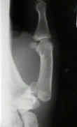
- Rolando's fracture
- Thumb Fractures/Dislocations
- X-ray Studies
- Discussion:
- most frequent of all thumb frx;
- described in 1882 by Dr. Edward Bennet;
- it is a frx dislocation, intra-articular frx at base of carpometacarpal joint of the thumb;
- involves an oblique intraarticular metacarpal frx (known as the palmar beak fragment) which remains
attached to the palmar beak ligament;
- anatomy of CMC joint
- mechanism of frx:
- results from axial blow directed against the partially flexed metacarpal; (ie. from fist fights)
- frx starts at ulnar base of thumb metacarpal;
- palmar ulnar aspect of thumb is normally stabilized by strong ligaments;
- disruption of the ulnar fragment destabilizes thumb;
- volar frx fragment remains attached to CMC by volar anterior oblique lig;
- anterior oblique ligament anchors volar lip of metacarpal to tubercle of the trapezium;
- hence, small volar lip fragment remains attached to anterior oblique ligament which is attached to trapezium;
- distal metacarpal fragment (containing most of articular surface) is displaced proximally, radially, & dorsally by pull of APL;
- displaced metacarpal is also rotated in supination by the pull of APL;
- metacarpal head is also displaced into palm by pull of ADP;

- Radiographs:
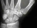
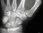
- oblique frx line with a triangluar fragment at ulnar base of metacarpal;
- triangular fragment remains attached to trapezium w/ proximal displacement of the metacarpal;
- note size of the volar lip fragment and the amount of displacement of shaft;
- Prognositic Features:
- location and displacement of the fracture;
- extent of crush or impaction at the metacarpal;
- presence or absence of shearing or impaction injury to radial side of articular surface of the trapezium;
- Reduction:
- the metacarpal shaft is displaced dorsally and radial direction due to the force of the abductor pollicis longus and adductor pollicis;
- reduction is accomplished w/ longitudinal traction on end of thumb, in addition to abduction and extension of thumb metacarpal;
- thumb is pronated to bring it into opposition w/ non-displaced palmar fragment;
- because the thumb CMC joint is incongrouos, upto 2 mm of articular displacement is well tolerated in Bennet fractures;
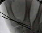
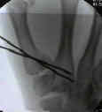
- consider closed reduction and percutaneous pin fixation when there is less than 3 mm of displacement, when
the beak of the fragment involves less than 50% of the palmar slope of the metacarpal, and when the concave
dome of the metacarpal is maintained;
- use 0.45 inch K wires to maintain reduction but do not attempt to spear small volar lip fragment with the wires;
- pins stabilize first metacarpal to trapezium or second metacarpal;
- may accept slight joint incongruity;
- if reduction not possible ORIF w/ AO cortical screw;
- spica cast for 4-6 weeks;
- Open Reduction:
- consider open reduction and internal fixation when there is more than 3 mm of fracture displacement;
- 2.5 cm transverse incision is made over radial base of thumb metacarpal;
- dorsal sensory branches of radial nerve are identified & protected;
- EPB & APL are identified and retracted;
- radial artery is protected and retracted ulnarly;
- by traction of thumb metacarpal, trapezial frx is visualized & reduced;
- frx reduction is provisionally secured w/ K wire;
- implants:
- if only 0.028-inch Kirschner Wires are used to secure trapezial frx, additional immobilization is required for six weeks;
- if radial fragment is of adequate size, 2.0-mm or 2.7 mm cortical lag screw is used;
- threaded or non threaded K wires (small sized fragments)
- 2.7 mm T or L plates
- 2.0 mm condylar plate
Long-term patient-reported outcomes following Bennett’s fractures
7-year follow-up after open reduction and internal screw fixation in Bennett fractures
Treatment of Bennett, Rolando, and vertical intraarticular trapezial fractures.
Trapeziometacarpal-I--Symposium: The Classic: On Fracture of the Metacarpal Bone of the Thumb.
Functional cast immobilization of thumb metacarpophalangeal joint injuries.
Fractures at the base of the thumb: treatment with oblique traction.
Post-traumatic instability of the metacarpophalangeal joint of the thumb.
Fractures of the basal joint of the thumb.
A long-term study following Bennett's fracture.
Long-term evaluation of Bennett's fracture. A comparison between open and closed reduction.


